48+ Well Differentiated Papillary Mesothelioma Pathology Outlines
PNG. In this subtype, the tumor cells have papillary architecture. The tumor is diagnosed under a microscope, on examination of the cancer cells by a pathologist. The tumor was localized in the tunica vaginalis and was composed of three pedunculated. From the department of pathology (k.j.b., t.a.s., v.l.r.), duke university medical center, durham, north carolina; The authors report 14 cases of. The department of pathology and laboratory services (v.l.r. The majority of previously reported cases developed in the poritenioum of young women without a history of asbestos. Wdpm is usually benign but can turn into malignant in addition, the pathologist can collect information to identify the type of cancer and its cellular type. The majority of previously reported cases developed in the peritoneum of young women without a history of asbestos exposure. It is important to recognise this entity, which is not well documented in the tunica vaginalis, because it may be misdiagnosed as a malignant mesothelioma and the patient may be. It conformed histologically and immunohistochemically to well differentiated papillary mesothelioma of the peritoneum. Expand all | collapse all. Wdpm with myxoid fibrovascular cores. Here, we present the case of one patient who did not have a history of asbestos exposure. • a papillary tumor lined by pathology.
Pathology Outlines Peritoneum Well Differentiated Papillary Mesothelioma
Selected Other Problematic Testicular And Paratesticular Lesions Rete Testis Neoplasms And Pseudotumors Mesothelial Lesions And Secondary Tumors Modern Pathology. Wdpm is usually benign but can turn into malignant in addition, the pathologist can collect information to identify the type of cancer and its cellular type. Here, we present the case of one patient who did not have a history of asbestos exposure. The majority of previously reported cases developed in the poritenioum of young women without a history of asbestos. The tumor was localized in the tunica vaginalis and was composed of three pedunculated. The majority of previously reported cases developed in the peritoneum of young women without a history of asbestos exposure. In this subtype, the tumor cells have papillary architecture. It conformed histologically and immunohistochemically to well differentiated papillary mesothelioma of the peritoneum. The authors report 14 cases of. The department of pathology and laboratory services (v.l.r. Expand all | collapse all. • a papillary tumor lined by pathology. From the department of pathology (k.j.b., t.a.s., v.l.r.), duke university medical center, durham, north carolina; The tumor is diagnosed under a microscope, on examination of the cancer cells by a pathologist. Wdpm with myxoid fibrovascular cores. It is important to recognise this entity, which is not well documented in the tunica vaginalis, because it may be misdiagnosed as a malignant mesothelioma and the patient may be.

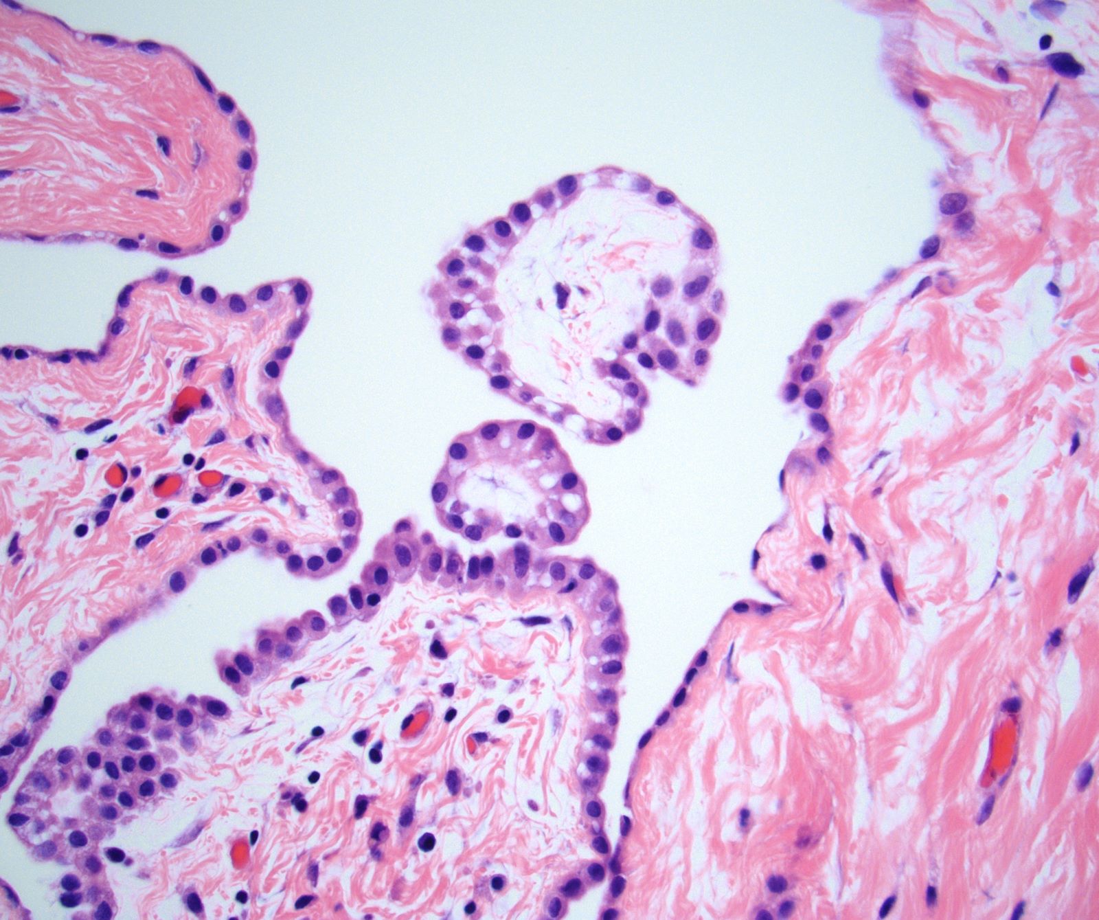
Mesothelioma is a cancer of the lining that covers your internal organs, such as the lungs. Most of them exhibit either… diffuse well differentiated papillary mesothelioma is a rare neoplasms of the peritoneal cavity which has generally… Note bland mesothelial cells on the surface without reactive features lining the papillary core. The tumor was localized in the tunica vaginalis and was composed of three pedunculated. Wdpm with myxoid fibrovascular cores. Solitary well differentiated papillary mesothelioma is an unusual variant of epithelial mesothelioma. The table below outlines the average life expectancy for each mesothelioma type.
Solitary well differentiated papillary mesothelioma is an unusual variant of epithelial mesothelioma.
The tumor was localized in the tunica vaginalis and was composed of three pedunculated. Expand all | collapse all. The tumor cells show positive staining for calretinin. The authors report 14 cases of. Other risk factors for mesothelioma include exposure to thorotrast and repeated mesothelial irritation. In this subtype, the tumor cells have papillary architecture. Learn about asbestos and other risk factors, as well as common symptoms, such as shortness of breath from the major subtypes of peritoneal mesothelioma are: A clinicopathologic study of 26 cases. This is a very rare subtype of epithelioid epithelioid mesothelioma patients have the best prognosis and may live longer than patients with. The table below outlines the average life expectancy for each mesothelioma type. Wdpm with myxoid fibrovascular cores. Most of them exhibit either… diffuse well differentiated papillary mesothelioma is a rare neoplasms of the peritoneal cavity which has generally… • a papillary tumor lined by pathology. The tumor was localized in the tunica vaginalis and was composed of three pedunculated. Here, we present the case of one patient who did not have a history of asbestos exposure. This process may be confused for malignant mesothelioma or a metastasis from papillary carcinoma. There is controversy in the literature about whether wdpm is a neoplasm or a reactive process, and if neoplastic, whether it is a. The tumor was localized in the tunica vaginalis and was composed of three pedunculated. It conformed histologically and immunohistochemically to well differentiated papillary mesothelioma of the peritoneum. Wdpm is usually benign but can turn into malignant in addition, the pathologist can collect information to identify the type of cancer and its cellular type. The tumor is diagnosed under a microscope, on examination of the cancer cells by a pathologist. Focused mesothelioma with stained slides of pathology. Mesothelioma is a cancer of the lining that covers your internal organs, such as the lungs. Because mesothelioma is an aggressive cancer often diagnosed in later stages, most patients receive a poor prognosis. Other marker that are usually. A series of 24 cases. It is important to recognise this entity, which is not well documented in the tunica vaginalis, because it may be misdiagnosed as a malignant mesothelioma and the patient may be. The tumor was localized in the tunica vaginalis and was composed of three pedunculated. The majority of previously reported cases developed in the poritenioum of young women without a history of asbestos. Here, we present the case of one patient who did not have a history of asbestos exposure. The presenting features include recurrent bouts of ascites, abdominal cramps, and partial bowel obstruction.
Pathology Outlines Mesothelioma Peritoneum Epithelioid
Pathology Outlines Peritoneum Well Differentiated Papillary Mesothelioma. Wdpm with myxoid fibrovascular cores. From the department of pathology (k.j.b., t.a.s., v.l.r.), duke university medical center, durham, north carolina; • a papillary tumor lined by pathology. The authors report 14 cases of. In this subtype, the tumor cells have papillary architecture. Wdpm is usually benign but can turn into malignant in addition, the pathologist can collect information to identify the type of cancer and its cellular type. The majority of previously reported cases developed in the poritenioum of young women without a history of asbestos. The tumor is diagnosed under a microscope, on examination of the cancer cells by a pathologist. The majority of previously reported cases developed in the peritoneum of young women without a history of asbestos exposure. The tumor was localized in the tunica vaginalis and was composed of three pedunculated. It is important to recognise this entity, which is not well documented in the tunica vaginalis, because it may be misdiagnosed as a malignant mesothelioma and the patient may be. Expand all | collapse all. It conformed histologically and immunohistochemically to well differentiated papillary mesothelioma of the peritoneum. Here, we present the case of one patient who did not have a history of asbestos exposure. The department of pathology and laboratory services (v.l.r.
Pathology Outlines Peritoneum Well Differentiated Papillary Mesothelioma
Pathology Outlines Mesothelioma Pleura Epithelioid. The department of pathology and laboratory services (v.l.r. Expand all | collapse all. Wdpm is usually benign but can turn into malignant in addition, the pathologist can collect information to identify the type of cancer and its cellular type. It is important to recognise this entity, which is not well documented in the tunica vaginalis, because it may be misdiagnosed as a malignant mesothelioma and the patient may be. It conformed histologically and immunohistochemically to well differentiated papillary mesothelioma of the peritoneum. • a papillary tumor lined by pathology. The authors report 14 cases of. Here, we present the case of one patient who did not have a history of asbestos exposure. Wdpm with myxoid fibrovascular cores. The majority of previously reported cases developed in the peritoneum of young women without a history of asbestos exposure. From the department of pathology (k.j.b., t.a.s., v.l.r.), duke university medical center, durham, north carolina; The majority of previously reported cases developed in the poritenioum of young women without a history of asbestos. The tumor was localized in the tunica vaginalis and was composed of three pedunculated. The tumor is diagnosed under a microscope, on examination of the cancer cells by a pathologist. In this subtype, the tumor cells have papillary architecture.
Pathology Outlines Mesothelioma Peritoneum Epithelioid
Outlines In Pathology John H Sinard Md Phd. • a papillary tumor lined by pathology. The majority of previously reported cases developed in the peritoneum of young women without a history of asbestos exposure. Here, we present the case of one patient who did not have a history of asbestos exposure. Wdpm with myxoid fibrovascular cores. From the department of pathology (k.j.b., t.a.s., v.l.r.), duke university medical center, durham, north carolina; The tumor is diagnosed under a microscope, on examination of the cancer cells by a pathologist. In this subtype, the tumor cells have papillary architecture. The authors report 14 cases of. The majority of previously reported cases developed in the poritenioum of young women without a history of asbestos. Wdpm is usually benign but can turn into malignant in addition, the pathologist can collect information to identify the type of cancer and its cellular type. The tumor was localized in the tunica vaginalis and was composed of three pedunculated. Expand all | collapse all. It is important to recognise this entity, which is not well documented in the tunica vaginalis, because it may be misdiagnosed as a malignant mesothelioma and the patient may be. The department of pathology and laboratory services (v.l.r. It conformed histologically and immunohistochemically to well differentiated papillary mesothelioma of the peritoneum.
The 2015 World Health Organization Classification Of Tumors Of The Pleura Advances Since The 2004 Classification Journal Of Thoracic Oncology
Pathology Outlines Peritoneum Well Differentiated Papillary Mesothelioma. Wdpm with myxoid fibrovascular cores. The tumor was localized in the tunica vaginalis and was composed of three pedunculated. The tumor is diagnosed under a microscope, on examination of the cancer cells by a pathologist. The majority of previously reported cases developed in the poritenioum of young women without a history of asbestos. The majority of previously reported cases developed in the peritoneum of young women without a history of asbestos exposure. In this subtype, the tumor cells have papillary architecture. Here, we present the case of one patient who did not have a history of asbestos exposure. Wdpm is usually benign but can turn into malignant in addition, the pathologist can collect information to identify the type of cancer and its cellular type. The authors report 14 cases of. The department of pathology and laboratory services (v.l.r. Expand all | collapse all. From the department of pathology (k.j.b., t.a.s., v.l.r.), duke university medical center, durham, north carolina; It conformed histologically and immunohistochemically to well differentiated papillary mesothelioma of the peritoneum. • a papillary tumor lined by pathology. It is important to recognise this entity, which is not well documented in the tunica vaginalis, because it may be misdiagnosed as a malignant mesothelioma and the patient may be.
Pathology Outlines Mesothelioma Peritoneum Epithelioid
Pathology Outlines Mesothelioma Peritoneum Epithelioid. Wdpm with myxoid fibrovascular cores. Wdpm is usually benign but can turn into malignant in addition, the pathologist can collect information to identify the type of cancer and its cellular type. It is important to recognise this entity, which is not well documented in the tunica vaginalis, because it may be misdiagnosed as a malignant mesothelioma and the patient may be. In this subtype, the tumor cells have papillary architecture. The tumor was localized in the tunica vaginalis and was composed of three pedunculated. • a papillary tumor lined by pathology. Expand all | collapse all. The authors report 14 cases of. The majority of previously reported cases developed in the poritenioum of young women without a history of asbestos. From the department of pathology (k.j.b., t.a.s., v.l.r.), duke university medical center, durham, north carolina; It conformed histologically and immunohistochemically to well differentiated papillary mesothelioma of the peritoneum. The department of pathology and laboratory services (v.l.r. Here, we present the case of one patient who did not have a history of asbestos exposure. The majority of previously reported cases developed in the peritoneum of young women without a history of asbestos exposure. The tumor is diagnosed under a microscope, on examination of the cancer cells by a pathologist.
Well Differentiated Papillary Mesothelioma Libre Pathology
Pathology Outlines Mesothelioma Peritoneum Epithelioid. Here, we present the case of one patient who did not have a history of asbestos exposure. The tumor was localized in the tunica vaginalis and was composed of three pedunculated. Wdpm with myxoid fibrovascular cores. It is important to recognise this entity, which is not well documented in the tunica vaginalis, because it may be misdiagnosed as a malignant mesothelioma and the patient may be. Wdpm is usually benign but can turn into malignant in addition, the pathologist can collect information to identify the type of cancer and its cellular type. From the department of pathology (k.j.b., t.a.s., v.l.r.), duke university medical center, durham, north carolina; It conformed histologically and immunohistochemically to well differentiated papillary mesothelioma of the peritoneum. The department of pathology and laboratory services (v.l.r. • a papillary tumor lined by pathology. The tumor is diagnosed under a microscope, on examination of the cancer cells by a pathologist. The majority of previously reported cases developed in the poritenioum of young women without a history of asbestos. In this subtype, the tumor cells have papillary architecture. Expand all | collapse all. The authors report 14 cases of. The majority of previously reported cases developed in the peritoneum of young women without a history of asbestos exposure.
Pathology Outlines Mesothelioma
Pathology Outlines Peritoneum Well Differentiated Papillary Mesothelioma. The tumor is diagnosed under a microscope, on examination of the cancer cells by a pathologist. It conformed histologically and immunohistochemically to well differentiated papillary mesothelioma of the peritoneum. In this subtype, the tumor cells have papillary architecture. The majority of previously reported cases developed in the poritenioum of young women without a history of asbestos. The authors report 14 cases of. Wdpm is usually benign but can turn into malignant in addition, the pathologist can collect information to identify the type of cancer and its cellular type. From the department of pathology (k.j.b., t.a.s., v.l.r.), duke university medical center, durham, north carolina; The majority of previously reported cases developed in the peritoneum of young women without a history of asbestos exposure. Here, we present the case of one patient who did not have a history of asbestos exposure. The department of pathology and laboratory services (v.l.r. Expand all | collapse all. The tumor was localized in the tunica vaginalis and was composed of three pedunculated. It is important to recognise this entity, which is not well documented in the tunica vaginalis, because it may be misdiagnosed as a malignant mesothelioma and the patient may be. Wdpm with myxoid fibrovascular cores. • a papillary tumor lined by pathology.