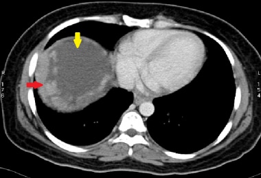
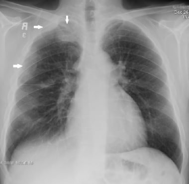
Pleural mesothelioma 90. Asbestosis is fibrosis of the lung caused by the presence of asbestos fibres in the lungs themselves it may have similar appearances to the fibrosis seen on the previous page.

Radiological Review Of Pleural Tumors Sureka B Thukral Bb Mittal
Pleural plaques are a benign entity do not lead to cancer or mesothelioma and their presence does not equate to the diagnosis of asbestosis note.

Pleural mesothelioma radiology. Pleural mesothelioma is a rare cancer that develops in the pleura a thin membrane of cells that line the lungs and chest wall. Tnm t tumor tx. Below is the international mesothelioma interest group tnm staging system.
Malignant pleural mesothelioma mpm is the most common primary tumor of pleura and its association with asbestos exposure has been well established. Pleural abnormalities can be subtle and it is important to check carefully around the edge of each lung where pleural abnormalities are usually more easily seen. There is a large pleural effusion present.
Computed tomography is the primary imaging modality used for the diagnosis and staging of mpm. Most tumors arise from the pleura and so this article will focus on pleural mesothelioma. Most tumors emerge from the pleura thus this article will concentrate on pleural mesothelioma.
No evidence of pr. Serum mesothelin related protein is a soluble form of mesothelin found to be elevated in 84 of patients with malignant mesothelioma and in less than 2 with other pulmonary or pleural diseases 10. Extension into interlobar fissures 40 86.
Given the presence of the mesothelium in different parts of the body mesothelioma can arise in various locations 17. This is best used as an adjunct to cyto pathologic and histopathologic examination in diagnosis of malignant mesothelioma. Pleural calcifications 20 circumferential encasement involvement of all pleural surfaces mediastinum pericardium fissures as late manifestation.
Mesothelioma also known as malignant mesothelioma is an aggressive malignant tumor of the mesothelium. The pleura only become visible when there is an abnormality present. Imaging plays an essential role in the evaluation of malignant pleural mesothelioma mpm.
A number of staging systems have been described for staging of malignant pleural mesothelioma. Some diseases of the pleura cause pleural thickening and others lead to fluid or air gathering in the pleural spaces. Pleural mesothelioma is the most common type of the disease accounting for 80 90 of all diagnoses.
Each year about 2500 people are diagnosed with pleural. Thick rind of irregular nodular malignant mesothelioma encases the left lung. Its incidence has been estimated at 22002500 cases per year in the united states 12.
Also known as malignant mesothelioma is a forceful threatening tumor of the mesothelium. Primary tumor cannot be assessed t0.
Pleural Effusion In Mesothelioma X Ray Stock Image C013 9674

Malignant Mesothelioma Imaging Overview Radiography Computed

Mesothelioma Radiology Case Radiopaedia Org

Iranian Journal Of Radiology Malignant Mesothelioma Versus

Malignant Mesothelioma Imaging Overview Radiography Computed

Radiological Review Of Pleural Tumors Sureka B Thukral Bb Mittal

