We studied the reactivity of malignant mesothelioma cells with tumor markers and the phenotypes of lymphocyte subsets in pleural effusions from 14 patients with malignant mesothelioma. The mesothelium is a membrane composed of simple squamous epithelium that forms the lining of several body cavities.

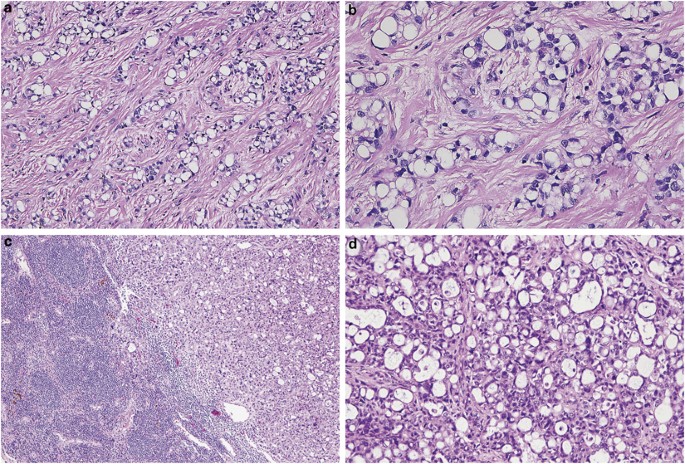
Mesothelioma With Signet Ring Cell Features Report Of 23 Cases Modern Pathology
The reaction pattern of mesothelioma cells was found to be cea negative leu m1 negative ema positive.

Mesothelioma histology microvilli. Mesothelial tissue also surrounds the male internal reproductive organs the tunica vaginalis testis and covers the internal reproductive organs of. Mesothelioma histology involves the study of cancerous mesothelial cells. Histology technicians make use of microscopes to visualize tissue and cells accurately.
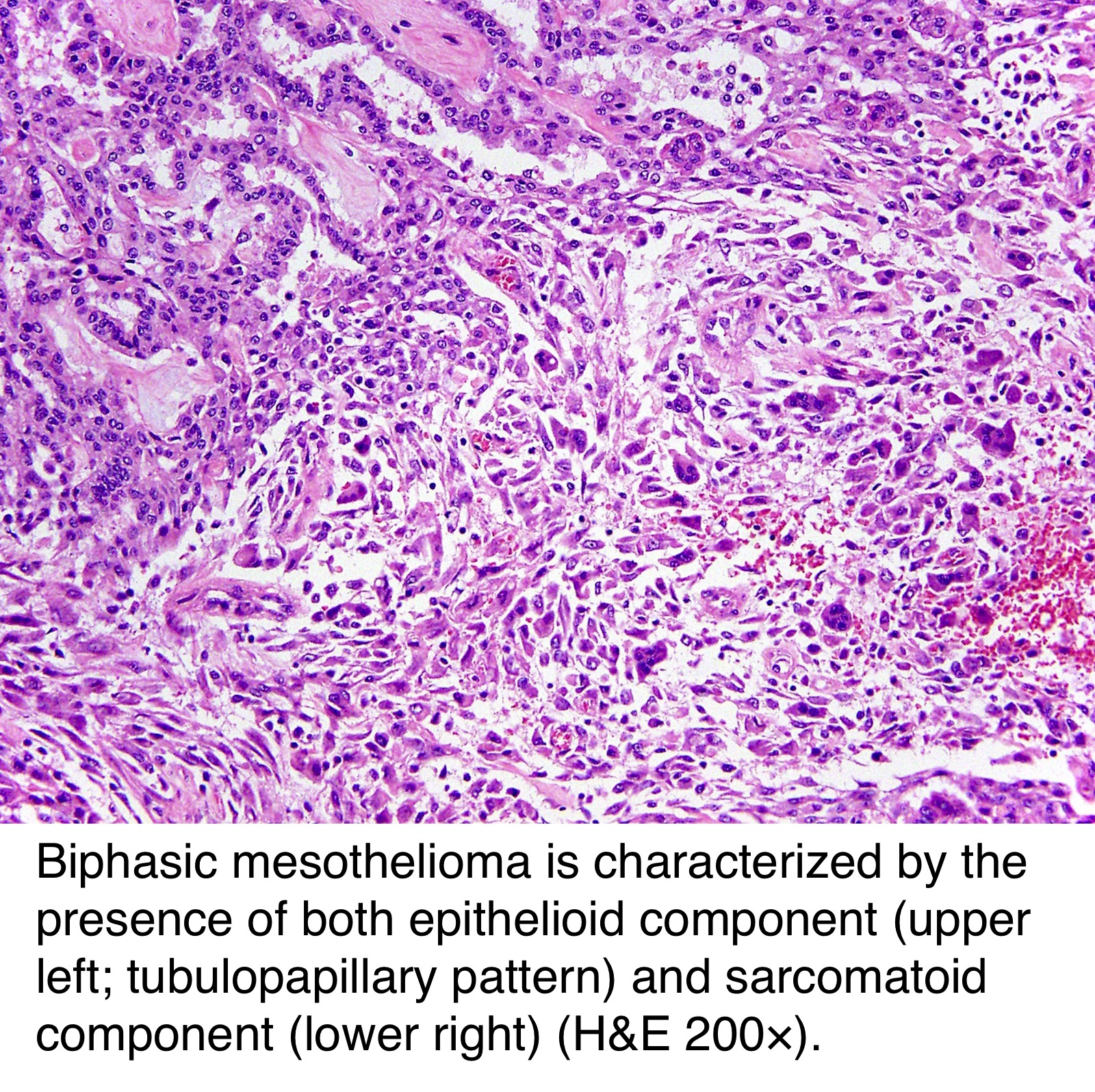
Doctors use these histological classifications to confirm the diagnosis. It is a branch of histopathology which studies diseased cells. With a distinctly round or cuboidal shape and elongated microvilli when viewed under a microscope.
Mesothelioma tumors typically present with three distinct histological abnormalities. Mesothelioma histology is the study of mesothelial cancer cells. Histology is an extended branch of biology in which cells and tissues are studied.
Mesothelioma histology an important tool used in the definitive diagnosis of disease is histology the microscopic examination of cellular anatomy. For identification of cell surface antigens with monoclonal antibodies the adhesive slide assay was used. Based on nine cases of mesotheliomas the sensitivity for long microvilli as a marker for mesothelioma is 889 and the specificity is 100.
Significance of studying mesothelium histology the study of histology of unnatural or diseased cells is termed histopathology. A fine needle aspiration biopsy of a pleural lesion resulted in a fragment of tissue composed of loosely aggregated tumor cells many of which have a considerable portion of the cell surface covered with microvilli m. The pleura thoracic cavity peritoneum abdominal cavity including the mesentery mediastinum and pericardium heart sac.
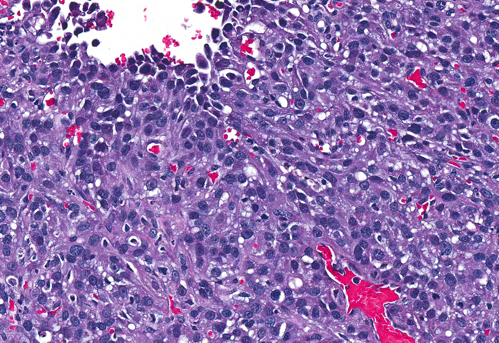
Histopathology of mesothelioma cells provide various insight to the illness and is a methods of accurate diagnosis. Histopathological studies can only be conducted with a certified and experienced medical professional. Hammar et al 21 reported four cases of mucicarmine positive mesothelioma in which tubular crystalloids were found to be associated with the microvilli in all four cases as well as in the.
The microvilli are profuse and rather than appearing to arise individually as straight projections. Epithelial cells have microvilli microscopic protrusions of the cell and clear structures called organelles within each cell. Long microvilli were observed in eight of nine cases of mesothelioma but not in 50 cases of mesothelial hyperplasia or 50 cases of nonmucinous pulmonary adenocarcinoma fig.

Malignant Mesothelioma Of Pleura Thoracic Pathology A Volume In The High Yield Pathology Series Expert Consult Online And Print 1st Edition

Pathology Outlines Peritoneal Malignant Mesothelioma

Conference 7 2017 Case 2 20171018

Early Detection Of Malignant Pleural Mesothelioma

Pdf Sarcomatoid Mesothelioma A Clinical Pathologic Correlation Of 326 Cases
Https Encrypted Tbn0 Gstatic Com Images Q Tbn 3aand9gcs9q5qyx6e27peswq P1qgcnejk5dfbyccejazpqk2e2yl7mico Usqp Cau
