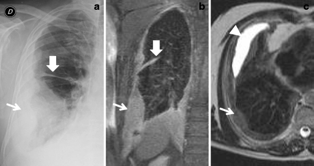
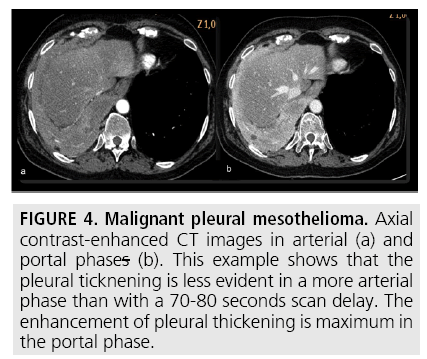
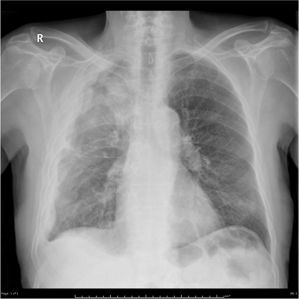
Often diagnosis is made after a patient has a chest x ray done for other reasons. As x ray technology developed further the framework for diagnosis and understanding the pathology of mesothelioma was established in 1931.

Plain Chest X Ray Findings In The Malignant Pleural Mesothelioma Download Table
The radiographer lines the machine up to make sure its in the right place.

Mesothelioma on cxr. A chest x ray is a medal imaging technique that is primarily used to diagnose injuries to bones. If you cant stand you can have it sitting or lying on the x ray couch. In these cases people may not even have symptoms.
You usually have a chest x ray standing up against the x ray machine. The most common area affected is the lining of the lungs and chest wall. The chest x ray therefore can be used to diagnose lung cancer pneumonia and mesothelioma cancer.
Sometimes the diagnosis is made after a chest x ray is taken for other reasons. Plaques may appear on the x ray image even though they are not causing symptoms. You must keep very still.
The earlier mesothelioma is discovered and operated on the better odds a patient has for survival. According to a 2019 study published in cancer imaging researchers are investigating the use of dynamic contrast enhanced mris in mesothelioma to better stage the cancer and to better predict a patients response to chemotherapy. Signs and symptoms of mesothelioma may.
Given the presence of the mesothelium in different parts of the body mesothelioma can arise in various locations 17. The term mesothelioma was coined in 1909 just a few years after the introduction of medical x ray imaging. Because of this symptoms typically affect breathing.
Less commonly the lining of the abdomen and rarely the sac surrounding the heart or the sac surrounding the testis may be affected. However may also be utilized to diagnose problems in a patients soft tissue including the lungs. The most common mesothelioma finding on radiographs is unilateral concentric plaque like or nodular pleural thickening.
Mesothelioma is a type of cancer that develops from the thin layer of tissue that covers many of the internal organs known as the mesothelium. Pleural effusions are common and may obscure the presence of the underlying pleural thickening. Here we present cxr from 64 year old hispanic male who was diagnosed with advanced mesothelioma.
Most people diagnosed with pleural plaques had no symptoms leading up to diagnosis. The lining of the lung is the most common site for the disease. The findings of ct scan are similar to those of chest x ray but are seen better and in more detail.
Mesothelioma also known as malignant mesothelioma is an aggressive malignant tumor of the mesothelium. Diagnosing pleural plaques and mesothelioma. Fewer than 5 of people exposed to asbestos develop mesothelioma.
The term was developed after thousands of autopsies numerous discoveries and significant research linked asbestos to a deadly form of cancer. For x rays of other areas of the body the best position is usually lying down on the x ray couch. Most tumors arise from the pleura and so this article will focus on pleural mesothelioma.
Pleural mesothelioma 90 covered in this article. In people with mesothelioma chest x ray may show signs of mesothelioma. However chest x ray has limited usefulness because the findings of mesothelioma on chest x ray are nonspecific and observed in other diseases as well.

Early Detection Of Malignant Pleural Mesothelioma

Imaging Characteristics Of Pleural Tumours Springerlink

Pleural Effusions Mesothelioma
Http Journal Chestnet Org Article S0012 3692 15 44250 3 Pdf

Diagnostic Imaging And Workup Of Malignant Pleural Mesothelioma

The Role Of Imaging In Malignant Pleural Mesothelioma Sciencedirect

