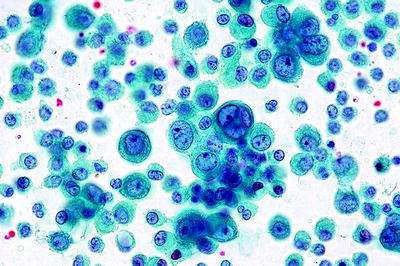
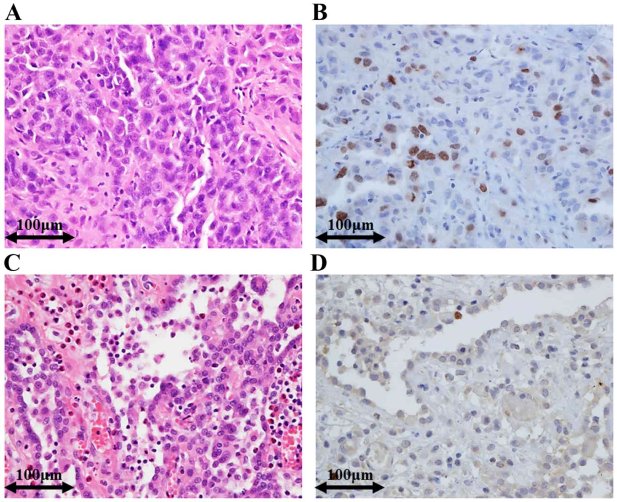
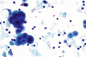
Of 217 cases circulated among all members of the uscanadian mesothelioma reference panel there was some disagreement about whether the process was benign or malignant in 22 of cases. Malignant mesothelioma 625 compared with reactive mesothelial proliferation 20 and adenocarcinoma 75.

Reactive Mesothelial Hyperplasia Springerlink
It deals with pericardial fluid peritoneal fluid and pleural fluid.

Mesothelioma vs reactive mesothelial cells cytology. In this study the authors investigated the utility of immunohistochemical ihc markers in making this distinction. On cell block sections a core of collagen and stromal cells surrounded by neoplastic cells is more commonly seen in mesothelioma than in adenocarcinoma whereas ring like structures with hollow cores are seen in some adenocarcinomas. Furthermore you may get more reliable information about the cell type from a histology report rather than just a cytology report.
Cytologic differential diagnosis among reactive mesothelial cells malignant mesothelioma and adenocarcinoma. Archival paraffin embedded cell blocks of pleural and peritoneal fluids from 52 patients with malignant mesothelioma mm and 64 patients with. 1 frank invasion is regarded as the most.
Reactive mesothelial cells in the backgrounds of adenocarcinomas were consistently negative for ecadherin. Mesothelial cells are often separated by slit like windows. Comparison of antibodies to hbme1 and calretinin for the detection of mesothelial cells in effusion cytology.
17 assessed ki 67 and other proliferation marker called repp86 and demonstrated that used in combination they are useful to discriminate between malignant mesothelioma and reactive mesothelial cells. The distinction of benign from malignant mesothelial proliferations in cytologic specimens can be problematic. Mesothelial cytopathology is a large part of cytopathology.
The morphological evaluation of cytological specimens from body cavity fluids presents difficulties in the differential diagnosis between benign reactive mesothelial rm cells and adenocarcinoma ac or malignant mesothelioma mm. Mesothelioma pathologists almost always request a tissue biopsy following a mesothelioma cytology report. The distinction between reactive mesothelial hyperplasia mh and malignant mesothelioma mm may be very difficult based only on histologic and morphologic findings.
A uniform cell population that is not signicantly different from one another were seen in 100 of cases of malignant mesothelioma as well as in reactive mesothelial proliferation. The aim of our study was to investigate whether a panel of five dif. Most adenocarcinoma smears showed a pop.
Most pathologists are reluctant to make a definitive diagnosis solely on fluid samples. The article deals with cytopathology specimens from spaces lined with mesothelium ie. Mesotheliomas than in reactive mesothelial hyperplasia and better results obtained with mcm2 11 16.
Http Www Asl5 Liguria It Portals 0 Anatomiapatologica2015 20150924 Effusion Cytology Pdf
Http Handouts Uscap Org 2016 Cm06 Daci 1 Pdf

Effusion Cytology Clinician S Brief

Utility Of Survivin Bap1 And Ki 67 Immunohistochemistry In Distinguishing Epithelioid Mesothelioma From Reactive Mesothelial Hyperplasia

Reliability Of P 16 Calretinin And Claudin 4 Immunocytochemistry In Diagnostic Verification Of Effusion Cytology
Https Www Rcpath Org Asset Ed8cdd8d 8d04 4b82 Ad48d585e2f023be

Mesothelioma Vs Reactive Mesothelial Cells Cytology Creative Art
