If a patient is supine then a pleural effusion layers along the posterior aspect of the chest cavity and becomes difficult to see on a chest x ray. If the patient is upright when the x ray is taken then fluid will surround the lung base forming a meniscus a concave line obscuring the costophrenic angle and part or all of the hemidiaphragm.

Cxr Pneumothorax Pleural Thickening
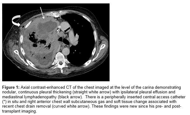
Some clinical studies show that when compared to an x ray a ct scan is better able to see pleural thickening asbestosis and pleural plaques.

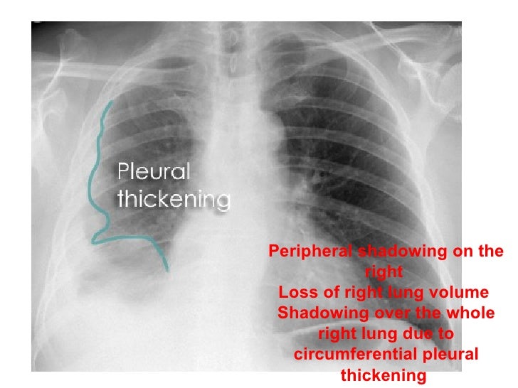
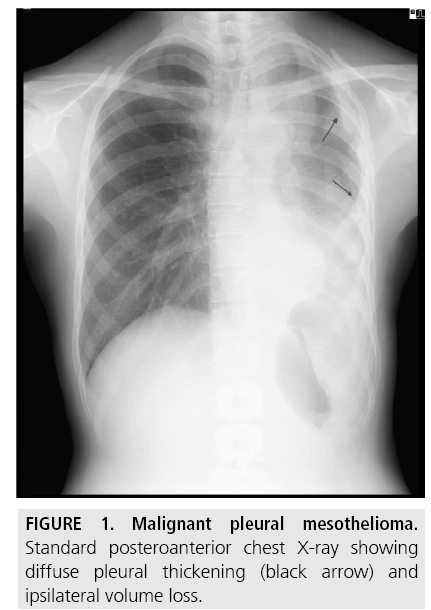
Pleural thickening mesothelioma x ray. The condition is most often diagnosed with a chest x ray but a diagnosis from a ct scan is better. A physician will check for physical symptoms such as altered breathing sounds. Given the presence of the mesothelium in different parts of the body mesothelioma can arise in various locations 17.
Often pleural thickening is diagnosed alongside its cause. Although it may not show everything necessary for diagnosis. A ct scan which is also used to diagnose asbestosis and pleural plaques can confirm the condition earlier.
Another finding is diffuse pleural thickening in the absence of pleural effusion. Healthy pleurae are not visible on an x ray but thickening at the very edges of the lungpleura may be. These scans are high resolution and allow doctors to distinguish between pleural thickening and pleural plaques.
This cancer is incurable but patients who are diagnosed early have a much greater life expectancy. An x ray can reveal thickening of the pleura but a ct scan is more likely to show thickening in better detail. Asbestos exposure can cause diffuse pleural thickening which results from lesions forming on the lung lining the pleura.
Methods used to detect and diagnose pleural thickening include. One of the initial signs of mesothelioma is a thickening of the lung or pleural thickening that can be seen on a chest x ray. People diagnosed with mesothelioma have aggressive cancer that is caused by asbestos exposure.
The most common way to diagnose pleural thickening is with x rays mris or ct scans. Ct scans also can see earlier signs of pleural thickening with scar tissue just 1 mm or 2 mm thick. Pleural thickening can be a sign of serious conditions such as malignant mesothelioma and lung cancer or it can be benign.
Pleural mesothelioma 90 covered in this article. Pleural thickening as well as pleural effusion and pleural classification as well as lymphonodi around hillus or mediastinum look more clearly and can be. You will learn can a chest x ray show mesothelioma and much more.
In terms of mesothelioma x ray findings may show a pleural mass on one side of the lung with pleural effusion or fluid. Pet and mri scans provide even further detail and may be used to differentiate pleural thickening from malignant mesothelioma. Mesothelioma also known as malignant mesothelioma is an aggressive malignant tumor of the mesothelium.
A ct or computed tomography scan gives a clearer picture of what is happening in the lungs and pleura. Most tumors arise from the pleura and so this article will focus on pleural mesothelioma. The pleural thickening in mesothelioma usually is in the part of lung basal or medio basal hemi 75 thorax3678910 chest ct scan in the mesothelioma shows more clearly than chest x ray.
A chest x ray is a typical first screening.

Pleural Tuberculosis A Key Differential Diagnosis For Pleural Thickening Even Without Obvious Risk Factors For Tuberculosis In A Low Incidence Setting Bmj Case Reports

Non Malignant Asbestos Related Diseases A Clinical View Rcp Journals
Https Www Resmedjournal Com Article S0954 6111 17 30033 1 Pdf

Obliteration Of Left Costophrenic Angle With Opaque Shadow Seen Along The Chest Wall Suggestive Of Lamellar Pleural Eff Mesothelioma Pleural Effusion Treatment

First Reported Finding Of A Malignant Pleural Mesothelioma In A Patient Post Liver Transplant Irish Medical Journal
Https Www Resmedjournal Com Article S0954 6111 17 30033 1 Pdf

