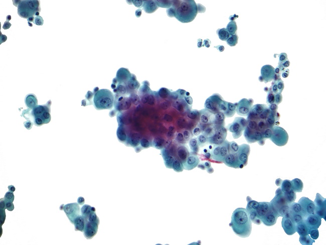
In this distinction pax8 expression has been regarded as a specific marker of serous carcinoma. For mesothelioma patients this fluid most commonly is either pleural effusion in pleural malignant mesothelioma patients or peritoneal effusion for those with mesothelioma of the peritoneum.
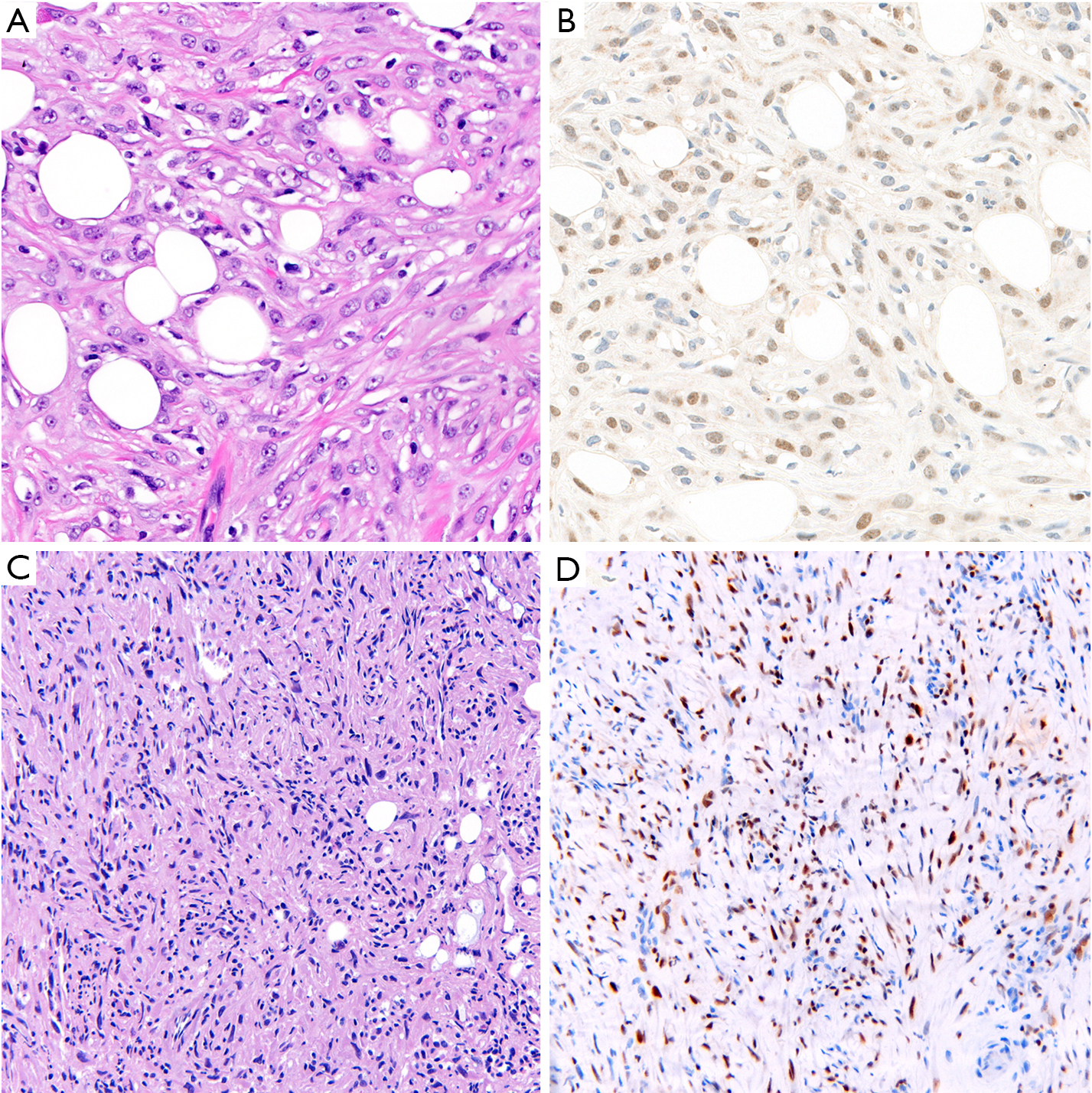
Application Of Immunohistochemistry In Diagnosis And Management Of Malignant Mesothelioma Chapel Translational Lung Cancer Research
Signs and symptoms of mesothelioma may.

Malignant mesothelioma cytology. The most common area affected is the lining of the lungs and chest wall. Mesothelioma specialists tend to have the best results when drawing cytology samples as they know exactly which parts of the body are most likely to collect mesothelioma cells. The tumour grows as multiple plaques and large nodules on the serosal surface.
Mesothelioma cytology or mesothelioma cytopathology is the study of cells for the presence of mesothelioma. Most patients have an effusion at the time of presentation. Siddiqui fernando schmitt andrew churg proceedings of the american society of cytopathology companion session at the 2019 united states and canadian academy of pathology annual meeting part 2.
In an effort to estimate the practice at other institutions a survey was disseminated regarding cytologic diagnosis of mm. Less commonly the lining of the abdomen and rarely the sac surrounding the heart or the sac surrounding the testis may be affected. Malignant mesothelioma arises most commonly in the pleura and rarely in the peritoneum.
It is a part of mesothelioma pathology which is the study of tissue or fluid to determine if this cancer exists. Malignant mesothelioma in cytology how far should we go. At the study institution northwestern university a primary diagnosis of mm is made on fluid cytology specimens.
The occurrence of this tumour is related to asbestos exposure. Follow the guidelines common sense clinicalimaging findings and remember behind every glass slide there is a human being and soon. Mesothelioma is a type of cancer that develops from the thin layer of tissue that covers many of the internal organs known as the mesothelium.
Mesothelioma cytology examines single cells often through analysis of bodily fluids. Distinguishing malignant peritoneal mesothelioma mpm from serous carcinoma involving the peritoneum remains a diagnostic challenge particularly in small biopsy and cytology specimens. Effusion cytology with focus on theranostics and diagnosis of malignant mesothelioma journal of the american society of cytopathology 10.
Never make a dx on cytology dx of mesothelioma on cytology piece of cake. Patients who suspect a mesothelioma diagnosis should therefore seek out the help and expertise of a qualified mesothelioma specialist. The diagnosis of malignant mesothelioma mm in effusion specimens is controversial.
In addition bap.
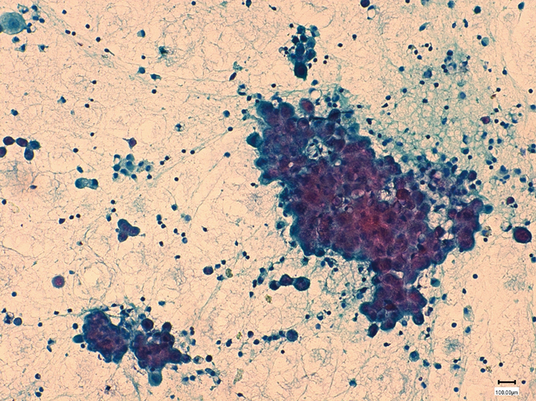
The Pathological And Molecular Diagnosis Of Malignant Pleural Mesothelioma A Literature Review Ali Journal Of Thoracic Disease

Malignant Mesothelioma Cytology

Cytomorphological Profile Of Neoplastic Effusions An Audit Of 10 Years With Emphasis On Uncommonly Encountered Malignancies Gupta S Sodhani P Jain S J Can Res Ther
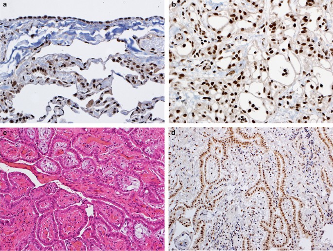
Bap1 Brca1 Associated Protein 1 Is A Highly Specific Marker For Differentiating Mesothelioma From Reactive Mesothelial Proliferations Modern Pathology
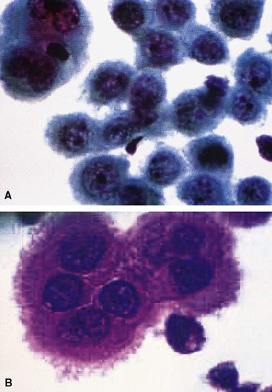
Malignant And Borderline Mesothelial Tumors Of The Pleura Thoracic Key
Https Encrypted Tbn0 Gstatic Com Images Q Tbn 3aand9gcsfu Mxmneu Gwnfgj0lkypoqodnbul8473zuq95ckzmrca Tnd Usqp Cau
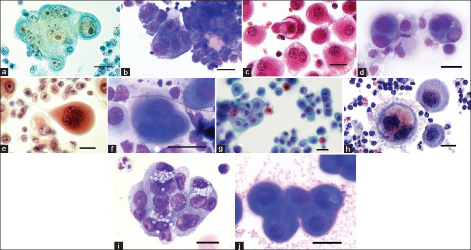
Guidelines For Cytopathologic Diagnosis Of Epithelioid And Mixed Type Malignant Mesothelioma Complementary Statement From The International Mesothelioma Interest Group Also Endorsed By The International Academy Of Cytology And The Papanicolaou Society Of
