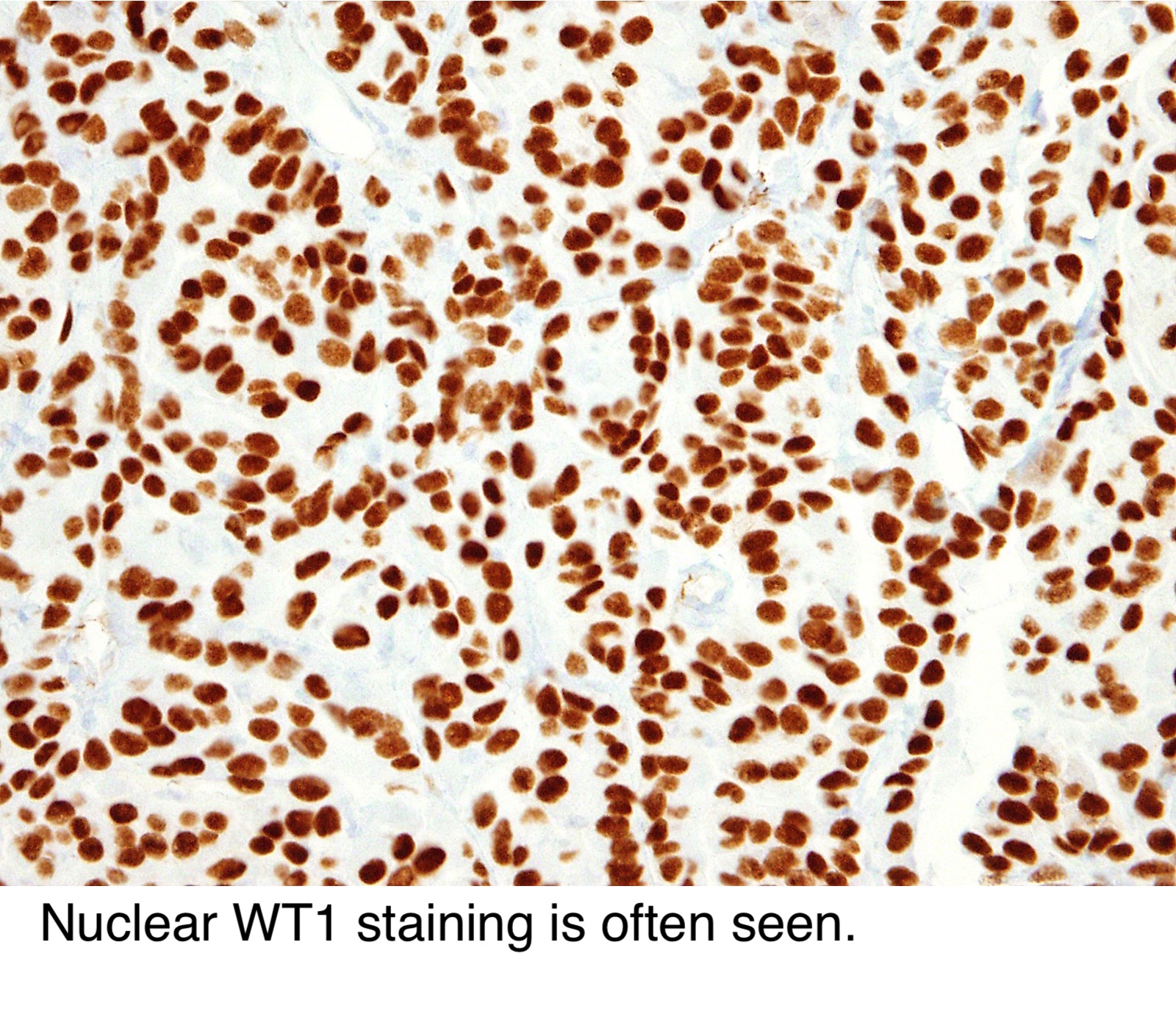
Differential diagnosis for elastofibroma. Wt1 is one of the most useful markers for identifying mesothelioma.
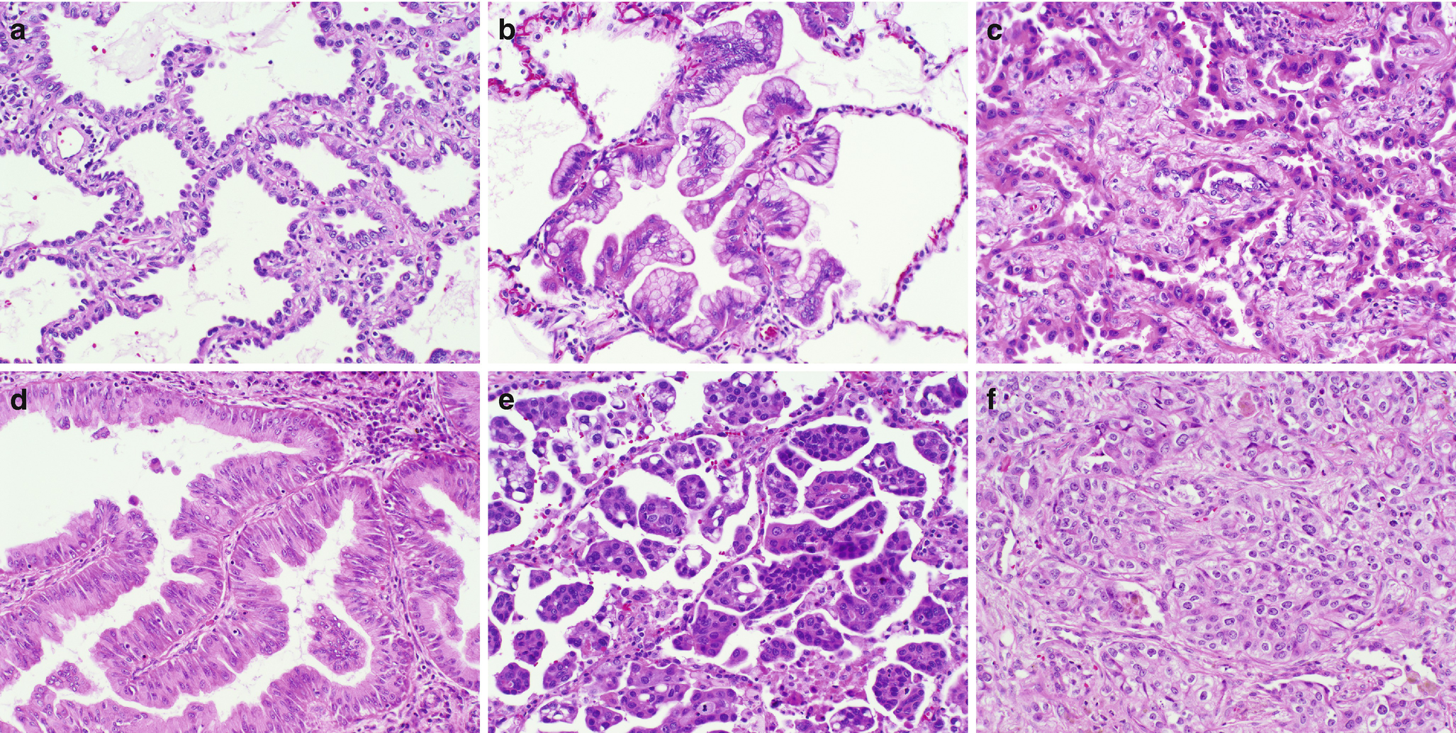
Lung Cancer Clinical Findings Pathology And Exposure Assessment Springerlink
16 17 however wt1 expression can also be detected in benign and malignant.
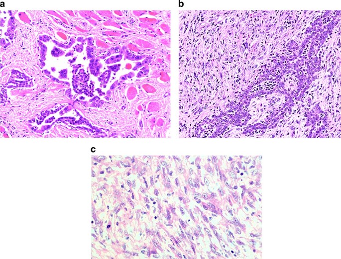
Wt1 mesothelioma pathology outlines. 1laboratory of pathology national cancer institute bethesda md 20892 usa. The occasional presence of signet ring cells may make it challenging to distinguish this disease from cancers of the lung. Malignant mesothelioma epithelioid variant 50 cm in greatest dimension surgical margins negative for tumor comment.
Mesothelioma histology or mesothelioma histopathology is the study of tissue for the presence of mesothelioma. Calretinin is an intracellular calcium binding ef hand protein of the calmodulin superfamily. Mesothelioma usually shows positivity for calretinin ck56 wt1 d2 40.
Wt1 may also play a role in making mesothelioma cells resistant to chemotherapy according to a 2017 study in pathology oncology research. After analyzing the results it is concluded that calretinin cytokeratin 56 and wt1 are the best positive markers for differentiating epithelioid malignant mesothelioma from pulmonary adenocarcinoma. The best discriminators among the antibodies considered to be negative markers for mesothelioma are cea moc 31 ber ep4 bg 8 and b723.
Wilms tumor gene product wt1 is a marker that is positively expressed in approximately 90 of primary ovarian carcinomas particularly in the serous subtype 1315 and has been used to distinguish carcinoma of ovarian origin from carcinoma with other primary sites. Immunohistochemical studies are essential for diagnosing mesothelioma and differentiating it from mimickers. Special studies for mesothelioma.
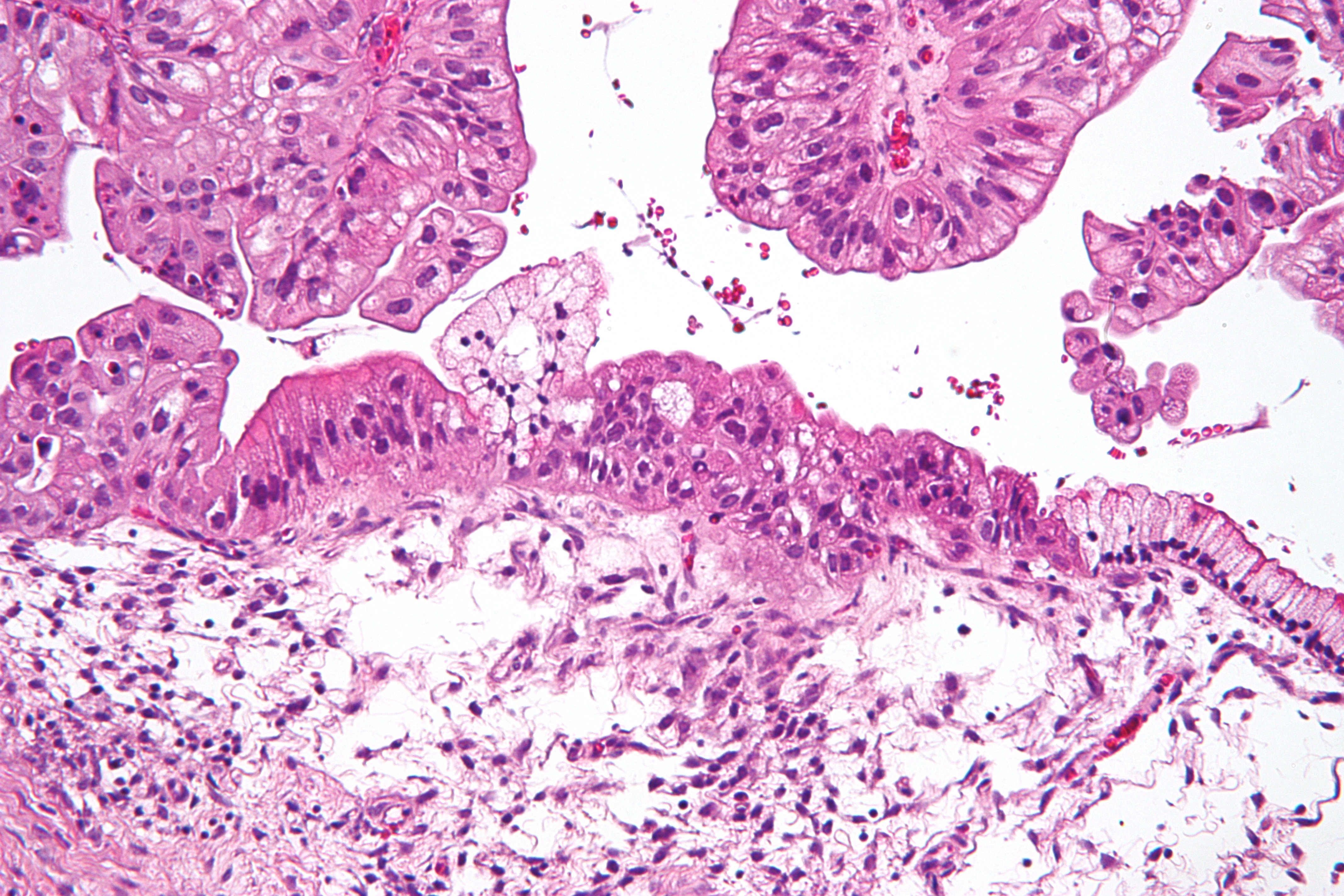
When researchers deactivated wt1 in laboratory mesothelioma cells their chemoresistance was suppressed. The tumor cells are positive for wt1 calretinin and d2 40 and are negative for claudin4 pax8 moc31 and cea. This process is part of mesothelioma pathology which involves examining either tissue or fluid to determine if this cancer exists in the body.
Histologically epithelial mesothelioma cells have polygonal ovoid or cuboidal cell shape. It plays a role in diverse cellular functions including message. This form of mesothelioma is comprised of cells which resemble the normal mesothelial cells in that they are arranged in a trabecular fashion.
Barak s1 wang z miettinen m.

Epithelioid Mesothelioma Treatment Symptoms Prognosis

Pathology Outlines Mesothelioma
Https Www Seap Es Documents 10157 1588536 The Challenge Of Cup Cadiz 2018 Janklos D024ee10 193d 42c7 8ff8 27d320c6d03a
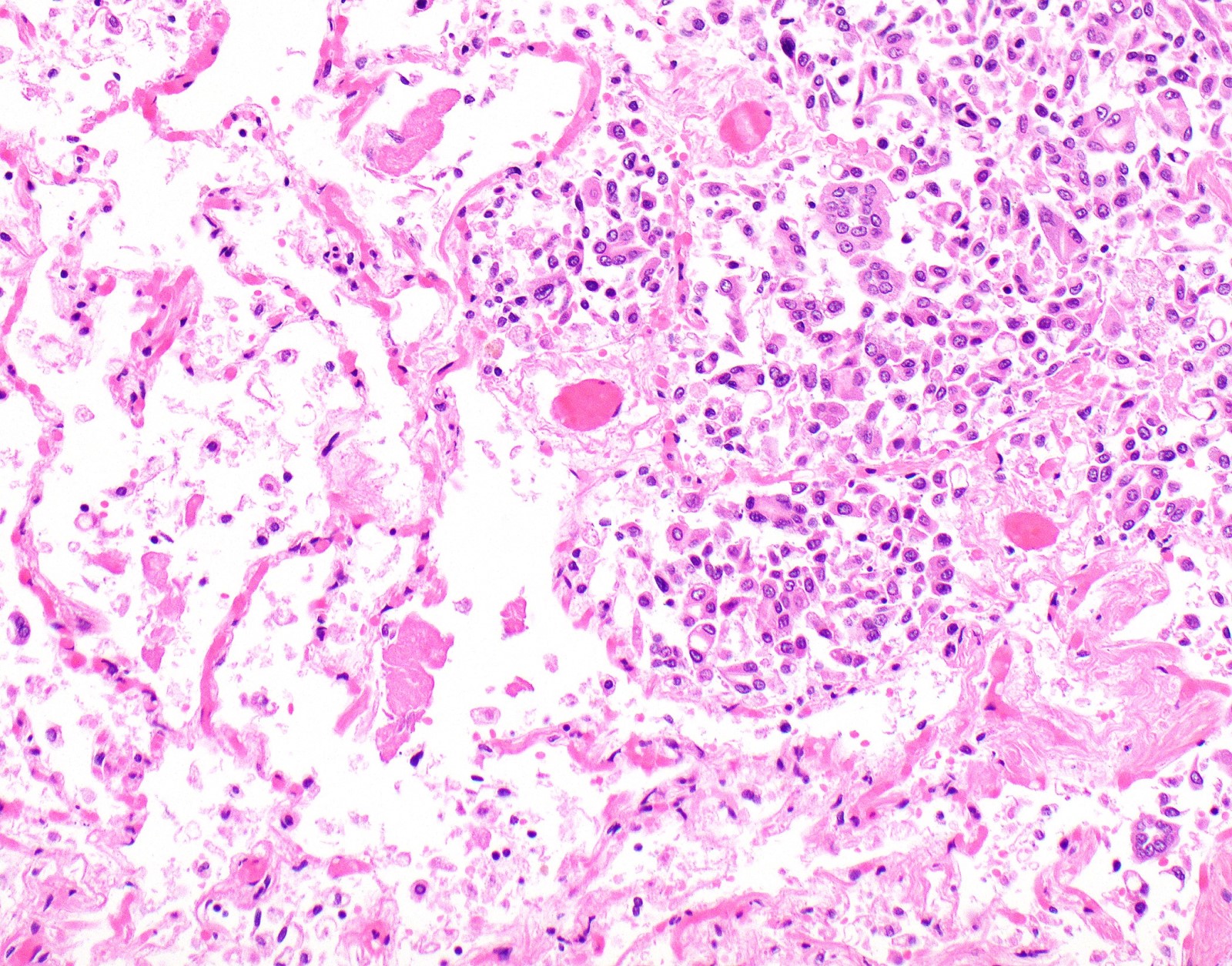
Pathology Outlines Diffuse Malignant Mesothelioma

Insert The Dark Side Of Mesothelioma Sciencedirect

Localized Malignant Mesothelioma An Unusual And Poorly Characterized Neoplasm Of Serosal Origin Best Current Evidence From The Literature And The International Mesothelioma Panel Modern Pathology
