40 80 of patients have a history of asbestos exposure. It generally progresses along that membrane resulting in breathing difficulties chest pain and fever.

An Overview Of How Asbestos Exposure Affects The Lung The Bmj
They often allow doctors to detect mesothelioma tumors including location sizing and potential spreading to aid in diagnosis and staging.

Mesothelioma ct chest. Go back to patient education resources learn about mesothelioma mesothelioma is a rare type of cancer that develops in the pleura a thin membrane that separates the lung from the chest wall. Imaging scans are often a first step in a mesothelioma diagnosis. If these tests indicate mesothelioma is likely the only way to confirm the disease is through a biopsy which involves the removal of fluid or tissue that will be examined under a microscope.
Once the mesothelioma doctor performs x rays on the patients lungs and if they show pleura or lung abnormalities the doctor may ask him to undergo a ct scan to deliver more precise results. Less commonly the lining of the abdomen and rarely the sac surrounding the heart or the sac surrounding the testis may be affected. It often results from a prior exposure to asbestos.
Mesothelioma is an uncommon entity and accounts for 5 28 of all malignancies that involve the pleura 17there is a strong association with exposure to asbestos fibers 10 risk during lifetime. Today x ray magnetic resonance imaging mri computed tomography ct and positron emission tomography pet nuclear scans are the four mesothelioma imaging tests used during the early stages of the diagnosis. Chest x ray showing a benign fibrous tumor of the pleura.
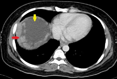
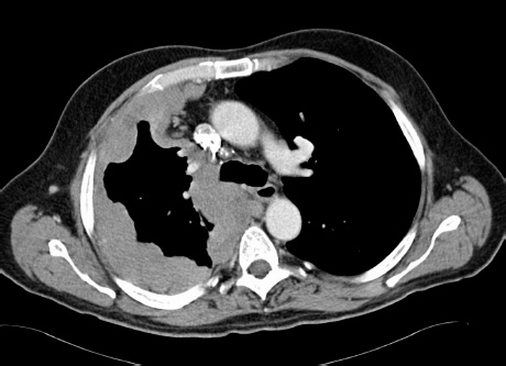
Ct scan of the chest demonstrates a large soft tissue mass in the right base which invades both the chest wall and the diaphragm and liver. A axial contrast enhanced chest ct scan shows a nodular right sided posterior pleural mass with associated calcification arrow a finding that is consistent with the patients known history of mesothelioma. X rays mri scans ct scans pet scans and ultrasounds are performed to detect abnormalities.
Unlike other asbestos related lung diseases it does not appear to be dose dependent 1. The most common area affected is the lining of the lungs and chest wall. Organizing fibrinous pleurisy epithelioid malignant mesothelioma sarcomatoid malignant mesothelioma and fibrous pleural plaques.
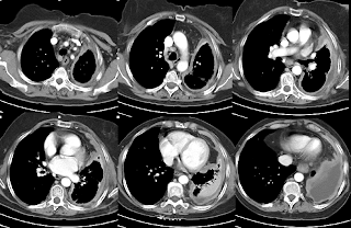
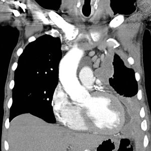
Typically patients with pleural mesothelioma receive a ct of the chest with contrast dye. These imaging tests are frequently used when making a mesothelioma diagnosis because they may help pinpoint the location of cancer in the body. He specializes in mesothelioma and cancers of the chest lungs and esophagus.
Computed tomography scans ct how it works electron beam cts mesothelioma chest scans. Mesothelioma is a type of cancer that develops from the thin layer of tissue that covers many of the internal organs known as the mesothelium. Ct scan revealing a pleural tumor.
He said the disease can be difficult to diagnose. Ct scans typically take 10 to 15 minutes to perform. Signs and symptoms of mesothelioma may.

Management Of Pleural Mesothelioma Thoracic Key

Malignant Mesothelioma Imaging Overview Radiography Computed

Ct2009 Diagnosis Malignant Mesothelioma With Contrast Ct Chest Images


