Ct scan if doctors suspect mesothelioma in their patients they may recommend various diagnostic tools including medical imaging procedures. Mesothelioma metastasis radiology ct scans are usually used for pleural mesothelioma and peritoneal mesothelioma but not for the pericardial type of the disease.

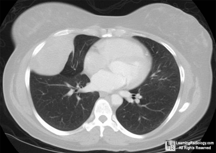
Learningradiology Localized Fibrous Tumor Pleura Solitary Benign Mesothelioma Pleural Fibroma Radiology
Staging of malignant pleural mesothelioma.

Ct scan mesothelioma radiology. It is generally considered too risky to use ct level radiation over the heart. Crossref medline google scholar. Comparison of ct and mr imaging.
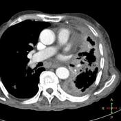
J digit imaging 2000. This image shows the extensive pleural thickening that is characteristic of mesothelioma effusion and reduction in the volume of the affected hemithorax. Malignant peritoneal mesothelioma is an uncommon primary tumor of the peritoneal lining.
It shares epidemiological and pathological features with but is less common than its pleural counterpart which is described in detail in the general article on mesothelioma. They often allow doctors to detect mesothelioma tumors including location sizing and potential spreading to aid in diagnosis and staging. Ct scans enable doctors to identify the stage of a tumor by exposing whether the tumor has spread to nearby tissues lymph nodes or to distant organs.
This imaging technology is less valuable in the diagnosis and staging of peritoneal mesothelioma but can be helpful in specific instances. The computed tomography scan is an important tool in this process because it can provide highly detailed information about the disease type location and metastasis. Other abdominal subtypes also discussed separately include.
Imaging scans are often a first step in a mesothelioma diagnosis. Resolution of pet scan images are relatively low hence the use of dual imaging combinations of pet and ct scans at most modern cancer centers. Initial chest radiography and chest computed tomography ct demonstrated circumferential lobulated pleural thickening involving the left lung with associated left lung volume loss figs 1 2whole body integrated positron emission tomography petct demonstrated circumferential pleural thickening surrounding the entire left lung with significant hypermetabolic.
21 heelan rt rusch vw begg cb panicek dm caravelli jf eisen c. X rays mri scans ct scans pet scans and ultrasounds are performed to detect abnormalities and potential causes of symptoms. Ct is most commonly used for imaging assessment of mesothelioma and sufficient for accurate staging of disease in most patients.
Pleural mass or nodular thickening of soft tissue attenuation tends to cause inward contraction of the hemithorax eg. Use of three dimensional spiral computed tomography imaging for staging and surgical planning of head and neck cancer. Computed tomography scan of a 58 year old patient with mesothelioma and shortness of breath.
Many doctors concur that ct scans provide the best imaging technology for scans of the abdomen and chest the areas most prone to the formation of mesothelioma.

Malignant Peritoneal Mesothelioma Radiology Reference Article Radiopaedia Org

Pleural Mesothelioma Dr Tinku Joseph

The Role Of Imaging In Malignant Pleural Mesothelioma An Update After The 2018 Bts Guidelines Clinical Radiology

Malignant Pleural Mesothelioma Evaluation With Ct Mr Imaging And Pet Radiographics

Malignant Peritoneal Mesothelioma Radiology Reference Article Radiopaedia Org
Https Encrypted Tbn0 Gstatic Com Images Q Tbn 3aand9gcrfu2kopu H3lo8g6sgjdfghewrsl9zt9czknshwv2eyugmcgbv Usqp Cau

Primary Peritoneal Mesothelioma Radiology Case Radiopaedia Org
