Mesothelioma histology or mesothelioma histopathology is the study of tissue for the presence of mesothelioma. Keratin 5 6.

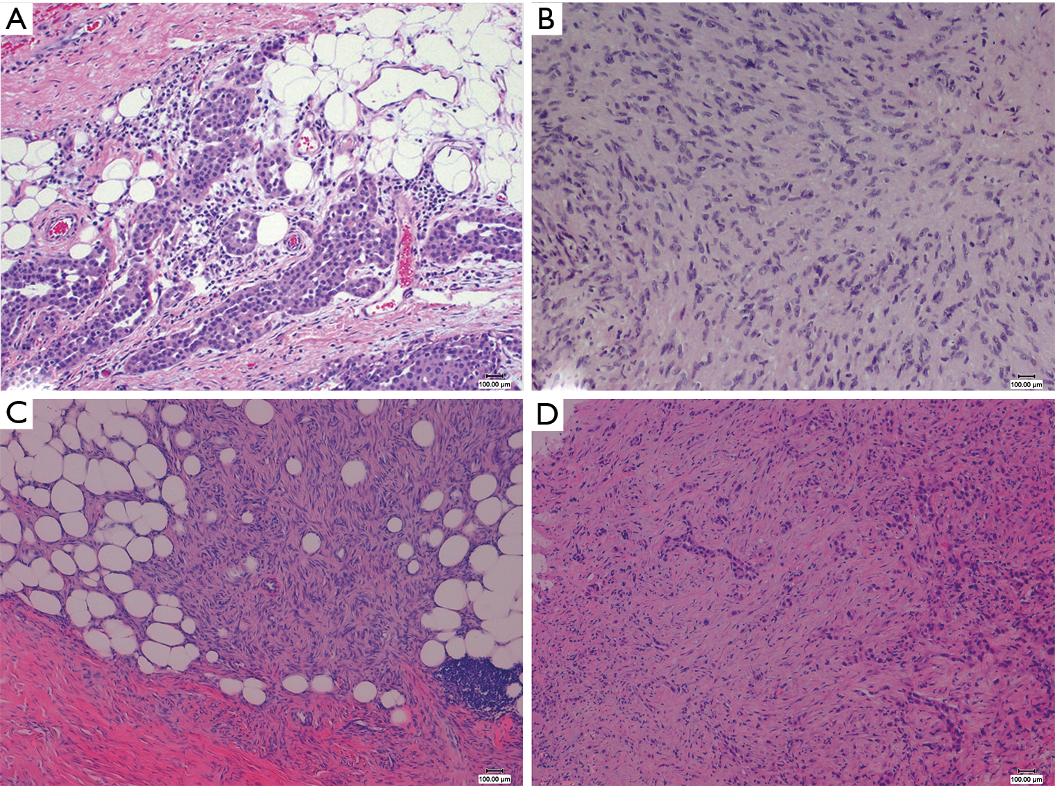
The Pathological And Molecular Diagnosis Of Malignant Pleural Mesothelioma A Literature Review Ali Journal Of Thoracic Disease
May be expressed in squamous cell carcinoma of lung and serous carcinoma synovial sarcoma and angiosarcoma.

Epithelioid mesothelioma histology. Expressed in epithelioid mesothelioma but negative in sarcomatoid mesothelioma podoplanin d2 40. Mesothelioma pathology focuses on understanding how mesothelial cells form spread and interact in the body. Specialists use it as a marker to distinguish epithelioid mesothelioma from lung adenocarcinoma.
This form of mesothelioma is comprised of cells which resemble the normal mesothelial cells in that they are arranged in a trabecular fashion. The occasional presence of signet ring cells may make it challenging to distinguish this disease from cancers of the lung. Mesothelioma histology involves two steps.
Epithelial mesothelioma cells can develop in the lining of the lungs abdomen or heart. D2 40 is a glycoprotein that shows an increased presence in many cancers including epithelioid mesothelioma. It has also been found useful in distinguishing epithelioid mesothelioma and squamous cell carcinoma.
Membranous and apical staining. Ncnn guidelines for treatment of cancer by site accessed 23 march 2018. Mutations in mesothelial cells caused by asbestos exposure create these variations.
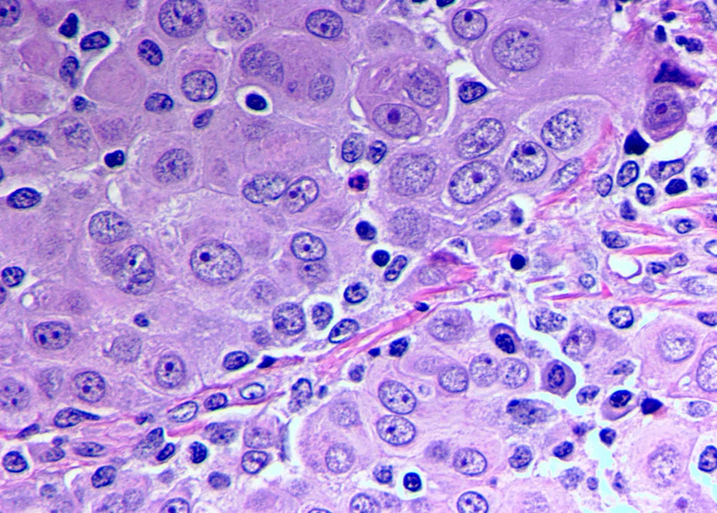
Sarcomatoid histology pure or mixed is poor prognostic factors after extrapleural pneumonectomy chemotherapy is recommended according to nccn guidelines national comprehensive cancer network. Epithelioid mesothelioma is caused by asbestos and is the most common type of the disease. Histologically epithelial mesothelioma cells have polygonal ovoid or cuboidal cell shape.
The tissue samples are sent to a laboratory where an experienced pathologist will review the samples and look for epithelioid sarcomatoid or biphasic mesothelioma cells. Pathologists study normal and diseased tissue to make an accurate diagnosissince mesothelioma is often mistaken for other diseases and cancers these cellular studies are vital in properly detecting mesothelioma before it becomes more advanced. This process is part of mesothelioma pathology which involves examining either tissue or fluid to determine if this cancer exists in the body.
The three major mesothelioma cell types are epithelioid sarcomatoid and biphasic. With treatment patients have an average life expectancy of one to two years. Doctors perform a tissue biopsy on a patient to collect tissue samples.
Treatment and prognosis are affected by a patients. The epithelial mesothelioma cell type makes up more than 50 of all cases.
Https Encrypted Tbn0 Gstatic Com Images Q Tbn 3aand9gcshqvyazp5jfs8 K H9mvknv80oo H Jnohlvuzuu4prxbww4 Q Usqp Cau
Https Mesotissue Org Sites Default Files 3 Hartman Meso Morphologic Subtypes Nmvb 2018 Pdf
Https Mesotissue Org Sites Default Files 3 Hartman Meso Morphologic Subtypes Nmvb 2018 Pdf

Early Detection Of Malignant Pleural Mesothelioma

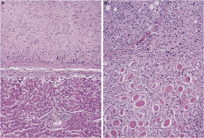
Sarcomatoid Mesothelioma A Clinical Pathologic Correlation Of 326 Cases Modern Pathology

Figure 2 From Reproducibility Of Histological Subtyping Of Malignant Pleural Mesothelioma Semantic Scholar

Epithelioid Mesothelioma Epithelial Cell Prognosis Treatment
