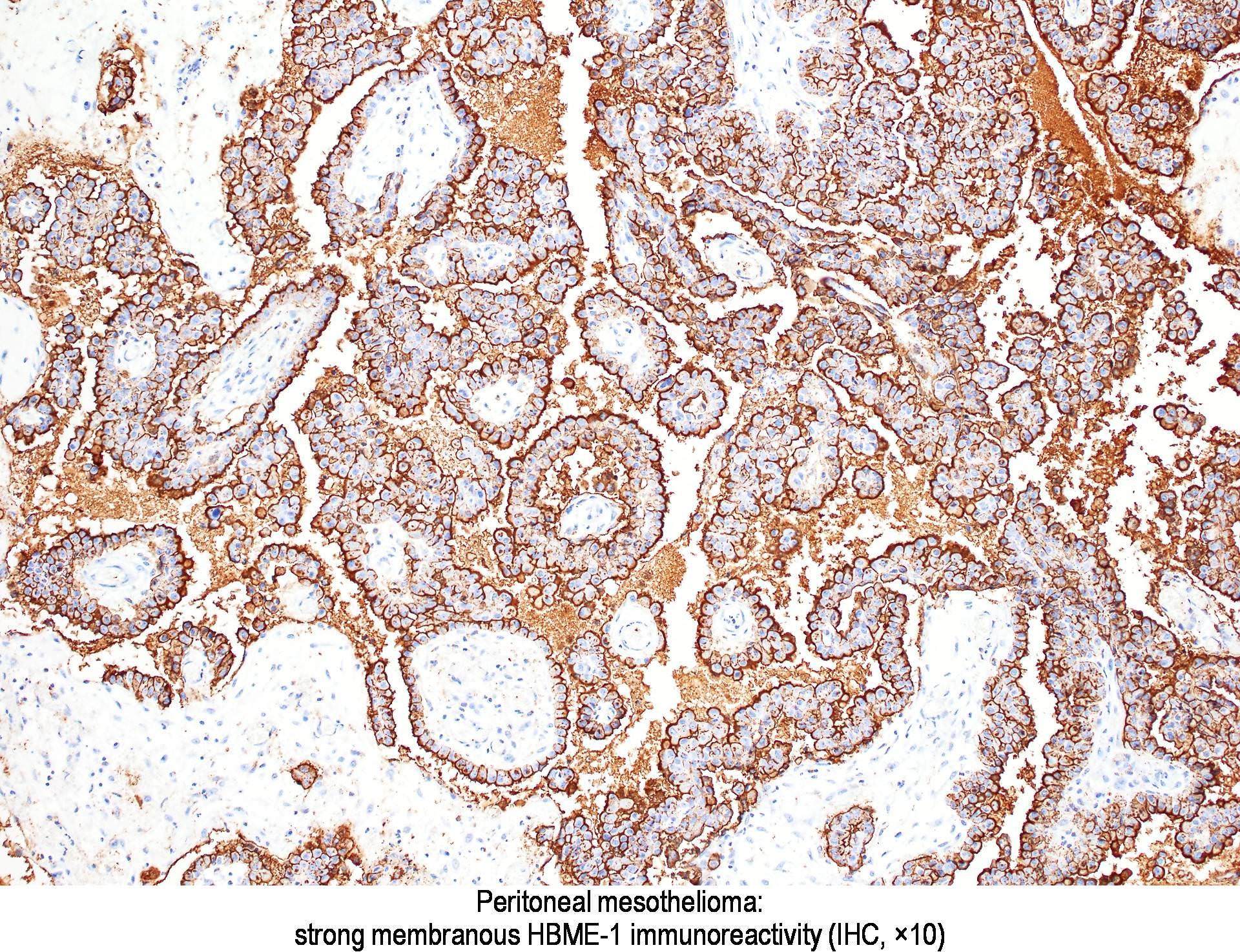
This is according to an article published in the journal of thoracic disease titled the use of electron microscopy for diagnosis of malignant pleural mesothelioma electron microscopy which was first developed in the 1930s uses a beam of accelerated electrons to magnify an image of a structure to a much higher degree of magnification. What is malignant mesothelioma electron microscopy.
Https Www Sciencedirect Com Science Article Pii S0031302516351078 Pdf Md5 E4dd5015175a552eff7a24d31ae34874 Pid 1 S2 0 S0031302516351078 Main Pdf
The use of electron microscopy for the diagnosis of malignant pleural mesothelioma transmission electron microscopy tem gave a great impulse to medical research.

Malignant mesothelioma electron microscopy. References prento p. Electron microscopy is a diagnostic tool using a type of microscopy which utilizes electrons to create an image. The nuclear pleomorphism is normally more extensive than seen in cytomorphology.
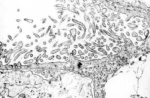
Epithelial malignant mesothelioma electron microscopy tremolite cleavage fragment renal cell carcinoma introduction while diagnosis of mesothelioma has become much easier in recent years with the advent of new immunochemical markers several cases still remain challenging. After its introduction for biological studies in 1931 several microscopic details from observation of animal and tumor cells by tem were published starting from 1953. A and b typically the apical surface of mm cells is covered by long slender microvilli completely devoid of any glycocalyx and malignancy is shown by the finding of neolumina that is.
103109019131232014960542 pmc free article pubmed cross ref. Histochem j 27 11. Glutaraldehyde for electron microscopy.
Malignant mesothelioma electron microscopy. Electron microscopy of malignant mesothelioma mm in effusions. Article january 2005.
But the method may be hampered by the fact that only slightly more than half of. Malignant mesothelioma mm is a neoplasm arising from mesothelial cells lining the pleural peritoneal and pericardial. An electron microscope is a powerful tool which allows for superior resolution of the specimen with the ability to magnify an object up to two million times.
Electron microscopy remains the gold standard for the diagnosis of epithelial malignant mesothelioma. A practical investigation of commercial glutaraldehydes and glutaraldehyde storage conditions. Transmission electron microscopy a high resolution method to explore cell characteristics can aid in the diagnosis of malignant pleural mesothelioma when examining cells found in the liquid surrounding the lungs.

Phenotypes And Karyotypes Of Huma Preview Related Info Mendeley

Pathology Outlines Diffuse Malignant Mesothelioma
A Patient With A Localized Malignant Pleural Mesothelioma And 15 Year Disease Free Survival Reig Oussedik Current Challenges In Thoracic Surgery

Genetic Predisposition To Fiber Carcinogenesis Causes A Mesothelioma Epidemic In Turkey Cancer Research
Https Link Springer Com Content Pdf 10 1007 2f0 387 28274 2 33 Pdf

Figure 3 From Metastatic Malignant Mesothelioma To The Tonsil Semantic Scholar

Malignant Mesothelioma And Other Mesothelial Proliferations Chapter 28 Modern Soft Tissue Pathology
