The diagnostic significance of the cytologic study of the fluid may be attributable to the fact that the cell population present in the sediment is representative of a. Numerous mesothelial cells are seen in this pleural fluid from a dog with a transudative effusion with concurrent diapedesis of red blood cells or hemorrhage.
Jcdr Adenocarcinoma Immunocytochemistry Reactive Mesothelial Cells Serous Effusions
Berger and stefan e.

Mesothelial cells in pleural fluid cytology. 20 of 119 of patients with malignancy involving the pleura but a negative pleural fluid cytology. An introduction to cytopathology is in the cytopathology article. Eighty five samples of mesothelial cells in pleural fluid from 76 patients with biopsy proven tuberculous pleurisy were examined cytologically.
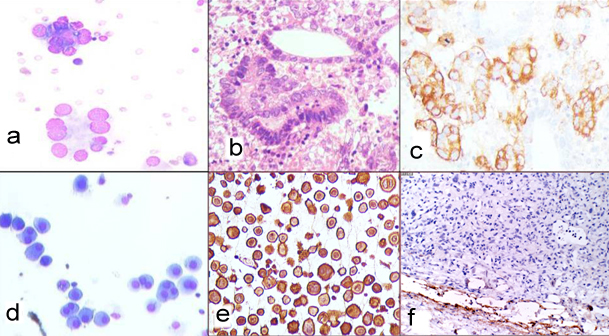
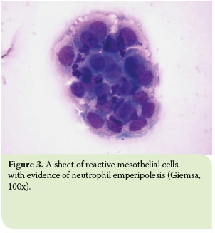
The morphological evaluation of cytological specimens from body cavity fluids presents difficulties in the differential diagnosis between benign reactive mesothelial rm cells and adenocarcinoma ac or malignant mesothelioma mm. Many reactive mesothelial cells were present in only 12 of samples analyzed. Actively dividing mesothelial cells can mimic an adenocarcinoma.
Pambuccian cytology of metastatic cervical squamous cell carcinoma in pleural fluid. Both non malignant and malignant causes of effusion can be identified by the relatively non invasive technique of pleural fluid cytologywith this basis the present study on cytology of pleural fluids was taken up. It deals with pericardial fluid peritoneal fluid and pleural fluid.
Gamez jose jessurun michael j. There are certain cells that line the pleura the thin double layered lining which covers the lungs chest wall and diaphragm which are known as mesothelial cellsother than the pleura mesothelial cells also form a lining around the heart pericardium and the internal surface of the abdomen peritoneum. Mesothelial cytopathology is a large part of cytopathology.
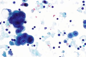
Mesothelial cells in pleural fluid. The aim of our study was to investigate whether a panel of five dif. The mesothelial cells have central round nuclei with a moderate amount of light purple cytoplasm and a corona or fringe to the cytoplasmic borders.
The article deals with cytopathology specimens from spaces lined with mesothelium ie. In contrast 653 of pleural fluid aspirates from a control group of patients with congestive heart failure contained. Report of a case confirmed by human papillomavirus typing diagnostic cytopathology 37 5 381 387 2009.
Cytology of mesothelial cells round with indistinct cell membranes and fuzzy borders single or clumped with so called windows between cells large round or elliptical nucleus often centrally placed with a single prominent nucleolus may contain two or multiple nuclei cytoplasm is green with papanicolaou and blue with mgg.
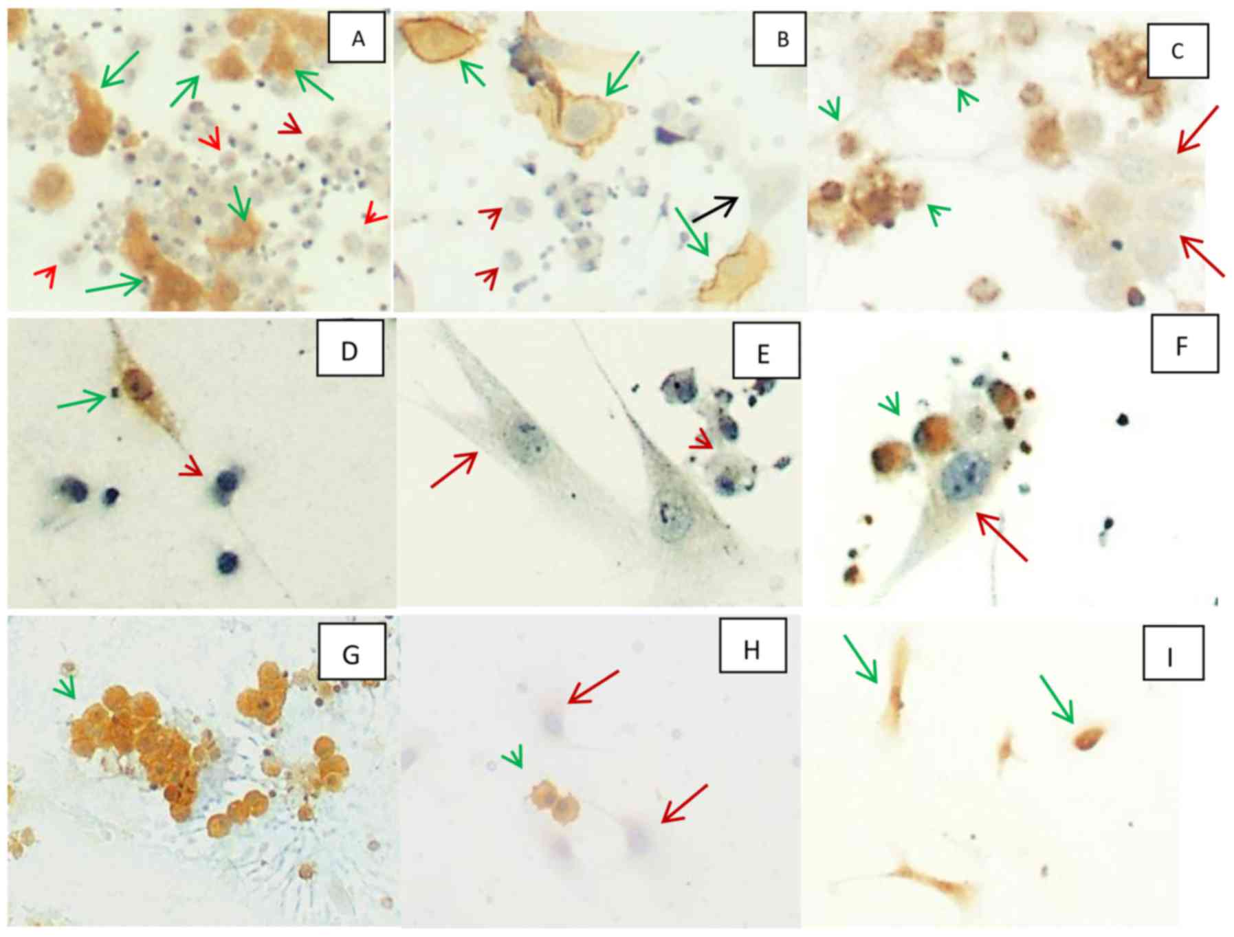
A Panel Of Markers For Identification Of Malignant And Non Malignant Cells In Culture From Effusions
Benign Pleural Fluid Cytology Acute Inflammatory Stock Photo Edit Now 552969733
![]()
Shutterstock Puzzlepix
Hjcam Iatrikh Zwwn Syntrofias Hellenic Journal Of Companion Animal Medicine Volume 6 Issue 1 2017 Pleural Effusion In The Cat A Focus On Laboratory Diagnosis

The Value Of Cytology And Pleural Biopsy In The Differential Diagnostic Of Nonspecific Pleural Effusions
Http Iap Ad Org Documents Archieve Congress 25thcongress Workshop First 20day Body 20fluids 20cytology Pdf

Utility Of Cell Block To Detect Malignancy In Fluid Cytology Adjunct Or Necessity Dey S Nag D Nandi A Bandyopadhyay R J Can Res Ther