The mesothelium is composed of an extensive monolayer of specialized cells mesothelial cells that line the bodys serous cavities and internal organs. In contrast 653 of pleural fluid aspirates from a control group of patients with congestive heart failure contained.

Pathology Glossary Pleural Effusions Draw It To Know It
Eighty five samples of mesothelial cells in pleural fluid from 76 patients with biopsy proven tuberculous pleurisy were examined cytologically.
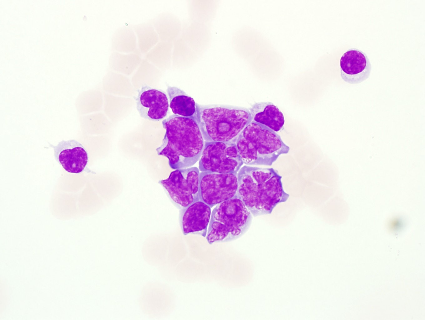
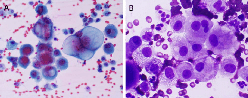
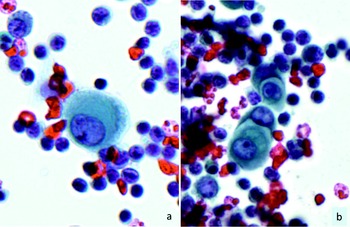
Mesothelial cells in pleural fluid images. Normally mesotheial cells present only along surface and not in underlying tissue. Pleural fluid cytological studies showed malignant cells in 33 of 43 patients with effusions due to tumor. Many reactive mesothelial cells were present in only 12 of samples analyzed.
Images hosted on other servers. Specific diagnoses benign eosinophilic pleuritis general. There are certain cells that line the pleura the thin double layered lining which covers the lungs chest wall and diaphragm which are known as mesothelial cellsother than the pleura mesothelial cells also form a lining around the heart pericardium and the internal surface of the abdomen peritoneum.
Of 31 exudative effusions with a lymphocytic predominance 30 were due either to tuberculosis or neoplasm. Pleural fluid right thoracentesis. Additional sampling should be considered within the clinical context.
Trauma with air in the pleural cavity. No tuberculous effusions had more than 1 mesothelial cells while most other effusions contained at least 5 mesothelial cells. Common cells present in pleural fluid include neutrophils lymphocytes monocytes mesothelial cells and red blood.
Epithelial or lining cells most commonly mesothelial cells1 the appearance and presentation of nucleated cells found in pleural fluid and whether they are considered commonbenign or abnormal is discussed below. Pleural fluid stock photos and images 319 matches. The main purpose of these cells is to produce a lubricating fluid that is released between layers providing a slippery non adhesive and protective surface to facilitate intracoelomic movement.
Negative for malignant cells. This has a large ddx. Reactive mesothelial cells present in a background of abundant lymphocytes.
65541208 mesothelial cell in pleural fluid. Add to likebox 31783093 illustration of human body respiratory system. Normal mesothelium in pelvic washings.
Mesothelial cells in pleural fluid. Find mesothelial cell pleural fluid stock images in hd and millions of other royalty free stock photos illustrations and vectors in the shutterstock collection. Thousands of new high quality pictures added every day.
This course is intended for laboratory professionals who have experience with peripheral blood morphology and basic experience with body fluid differential analysisthis tutorial will provide a review of normal and abnormal body fluid morphology utilizing wright giemsa stained cytospin preparations from cerebrospinal fluid csf pleural peritoneal and synovial fluids as. Mesothelial cells form conspicuous layer of regularly spaced bland cuboidal cells along pleural surface.
Http Www Cap Org Apps Docs Committees Hematology Educational Activities 2009 Cmb Pdf
Cytology Of Pleural And Peritoneal Lesions Chapter 5 Practical Pathology Of Serous Membranes
Https Encrypted Tbn0 Gstatic Com Images Q Tbn 3aand9gcr S D Awpannkakza 0ldk Epafkthta17xwyo8 N I 9tiksc Usqp Cau
A Mesothelial Cell Arrow In A Pleural Clinical Pathology College Of Veterinary Medicine Cornell University Facebook

Home
Https Encrypted Tbn0 Gstatic Com Images Q Tbn 3aand9gcqhjpudk4mltvqtva5hp2nrmoggfywdmzos5bx1le6l Sprigk Usqp Cau

The Value Of Cytology And Pleural Biopsy In The Differential Diagnostic Of Nonspecific Pleural Effusions