An introduction to cytopathology is in the cytopathology article. Add to likebox 31783093 illustration of human body respiratory system.
Https Encrypted Tbn0 Gstatic Com Images Q Tbn 3aand9gcqhjpudk4mltvqtva5hp2nrmoggfywdmzos5bx1le6l Sprigk Usqp Cau
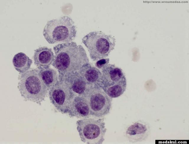
Clusters of 12 cells is unusual in simple hyperplasia binucleation multinucleation mitosis prominent nucleolus can be seen in benign proliferations two or more mesothelial cells are often separated by window or a narrow space benign mesothelial cells usually have characteristic skirt or halo at pale outer rim of cell.
Mesothelial cells in pleural fluid pictures. 65541208 mesothelial cell in pleural fluid. Thousands of new high quality pictures added every day. Mesothelial cells in pleural fluid eighty five samples of mesothelial cells in pleural fluid from 76 patients with biopsy proven tuberculous pleurisy were examined cytologically.
It deals with pericardial fluid peritoneal fluid and pleural fluid. Mesothelial cytopathology is a large part of cytopathology. Pleural fluid stock photos and images 319 matches.
Mesothelial cells are horizontally oriented venules are perpendicular oriented zonation of mesothelial cells highest cellularity near serosal surface trailing off toward chest wall no invasion no tumefactive growth j clin pathol 200659564. It is not uncommon for macrophages to be mixed in a ball reactive mesothelial cells. There are certain cells that line the pleura the thin double layered lining which covers the lungs chest wall and diaphragm which are known as mesothelial cellsother than the pleura mesothelial cells also form a lining around the heart pericardium and the internal surface of the abdomen peritoneum.
Of 31 exudative effusions with a lymphocytic predominance 30 were due either to tuberculosis or neoplasm. Distorted architecture of pleura in contrast benign pleura. Pleural fluid cytological studies showed malignant cells in 33 of 43 patients with effusions due to tumor.
No tuberculous effusions had more than 1 mesothelial cells while most other effusions contained at least 5 mesothelial cells. The article deals with cytopathology specimens from spaces lined with mesothelium ie. Mesothelial cells in pleural fluid.
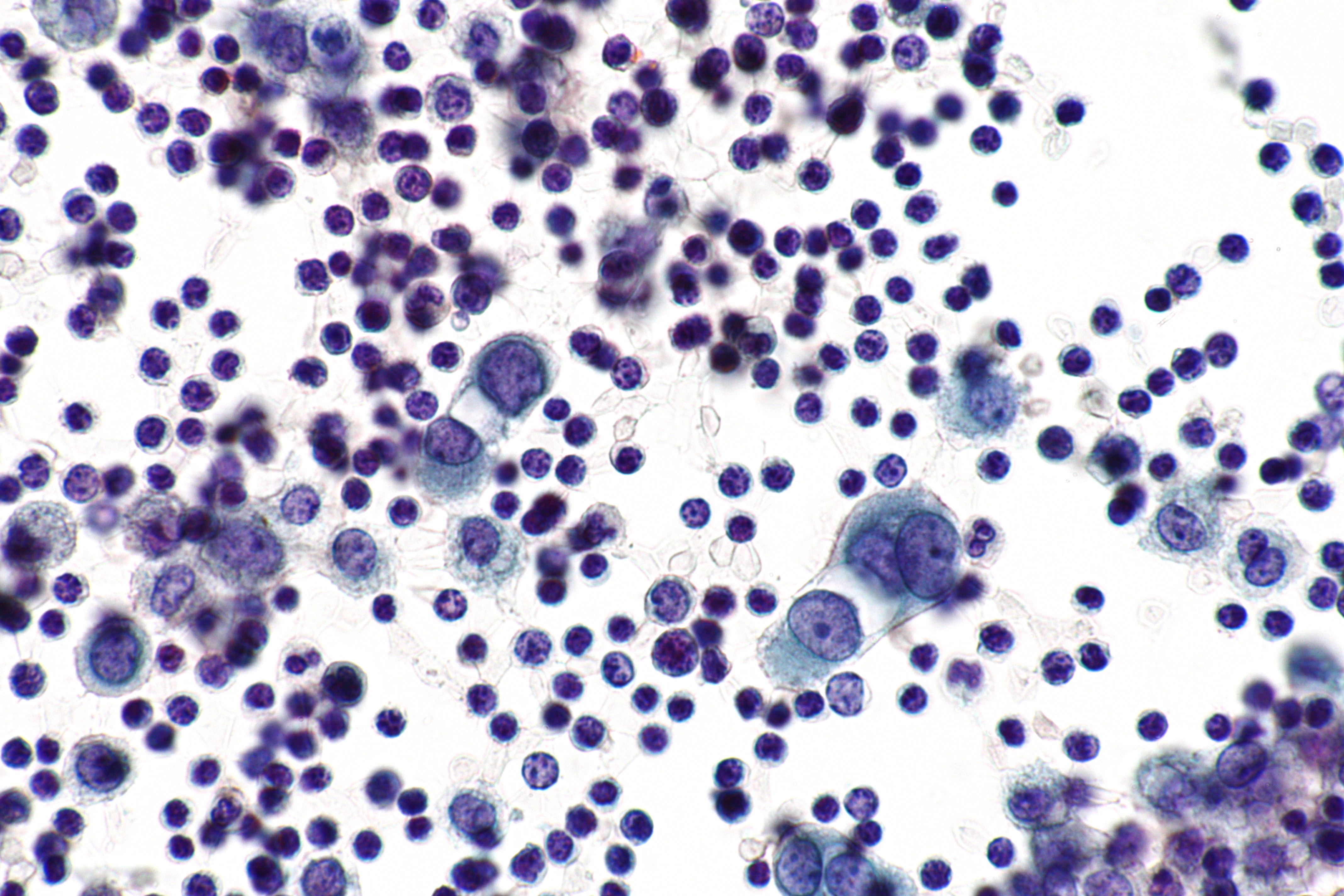
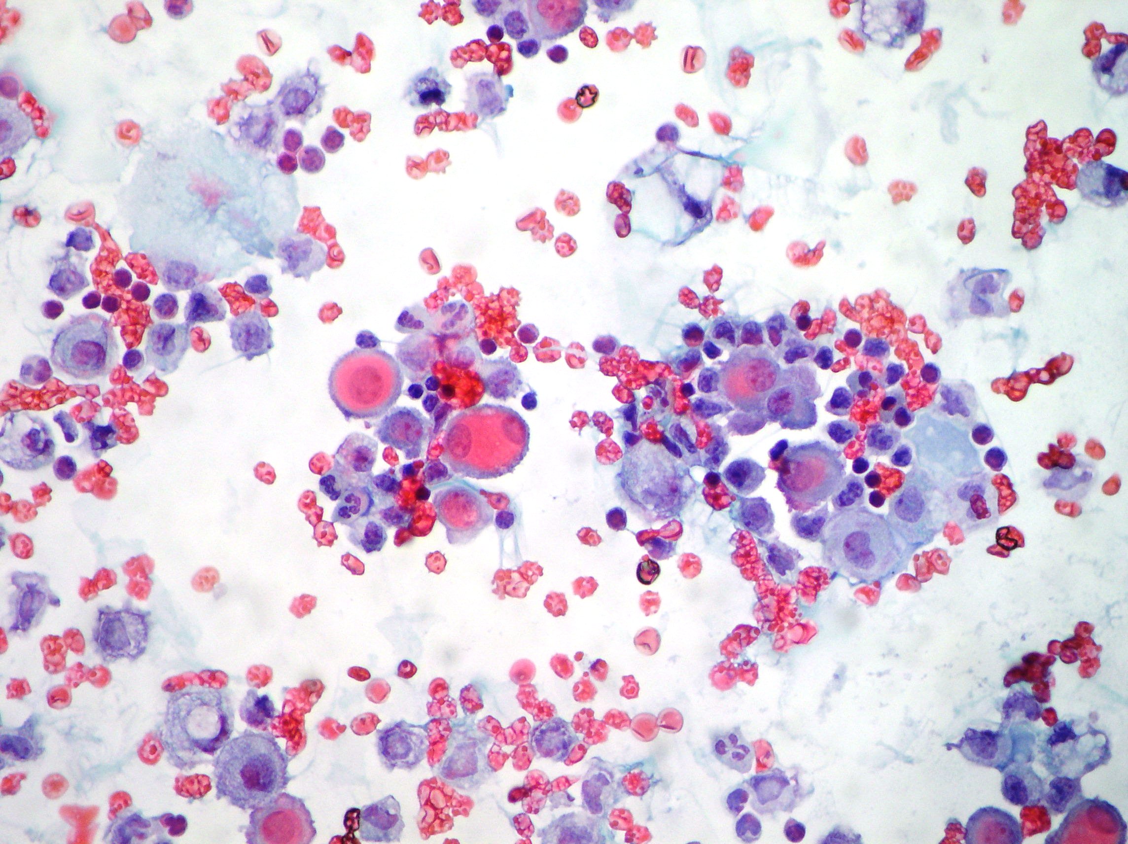
Many reactive mesothelial cells were present in only 12 of samples analyzed. Common cells present in pleural fluid include neutrophils lymphocytes monocytes mesothelial cells and red blood. Find mesothelial cell pleural fluid stock images in hd and millions of other royalty free stock photos illustrations and vectors in the shutterstock collection.
Picture of mesothelial cell in pleural fluid stock photo images and stock photography. Epithelial or lining cells most commonly mesothelial cells1 the appearance and presentation of nucleated cells found in pleural fluid and whether they are considered commonbenign or abnormal is discussed below.

Mesothelial Cytopathology Libre Pathology

Serous Effusions Basicmedical Key
File Benign Mesothelial Cells Pleural Fluid High Mag Jpg Wikimedia Commons
Effusions Cytopathology Cellnetpathology

Cytology Of Pleural And Peritoneal Lesions Chapter 5 Practical Pathology Of Serous Membranes
Cytology Of Pleural And Peritoneal Lesions Chapter 5 Practical Pathology Of Serous Membranes

Cytology Of Pleural And Peritoneal Lesions Chapter 5 Practical Pathology Of Serous Membranes