Tuberculous pleurisy is the most frequent extrapulmonary manifestation of tuberculosis. 643 and 3 514.
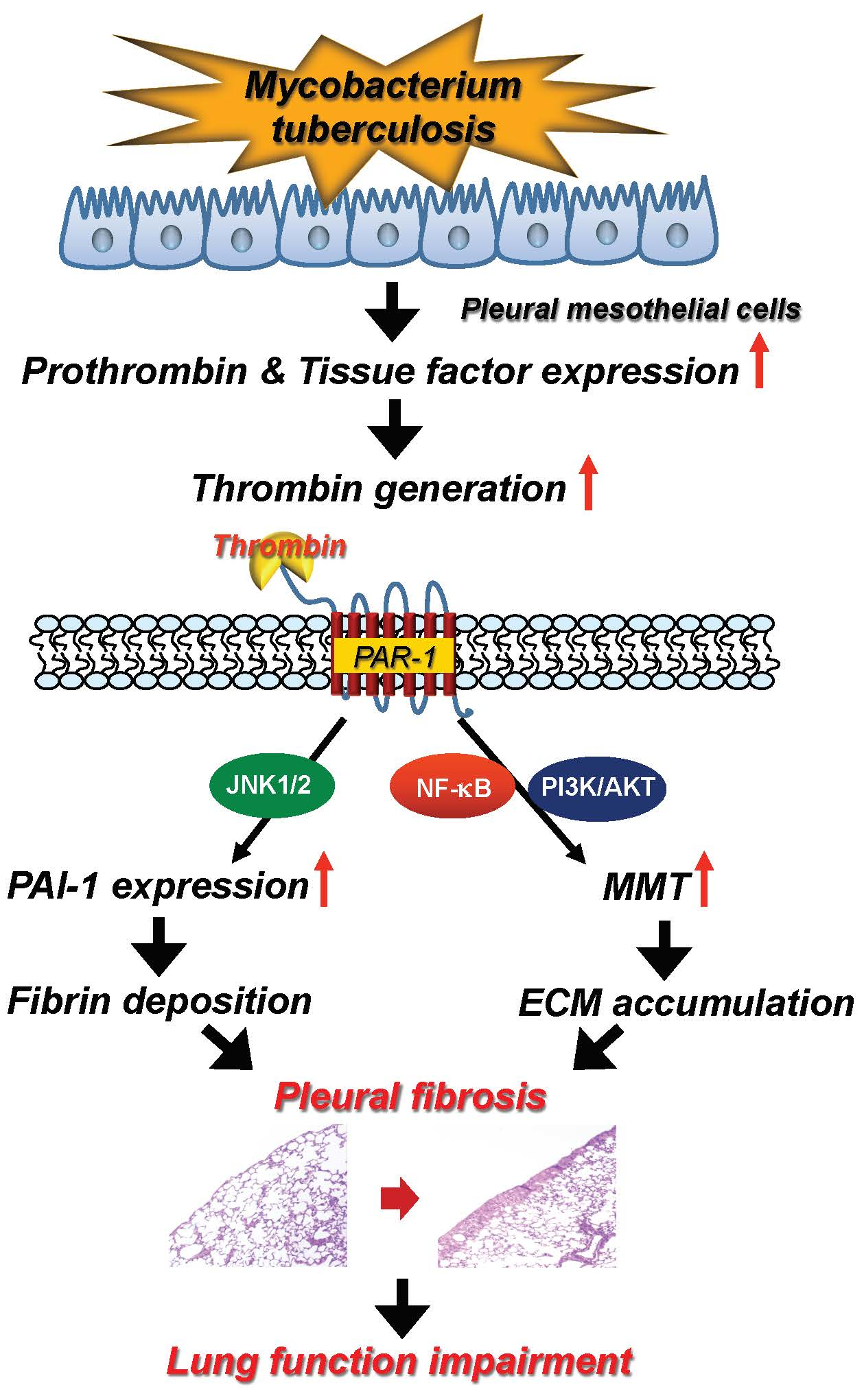
Ijms Free Full Text Thrombin Upregulates Pai 1 And Mesothelial Mesenchymal Transition Through Par 1 And Contributes To Tuberculous Pleural Fibrosis Html
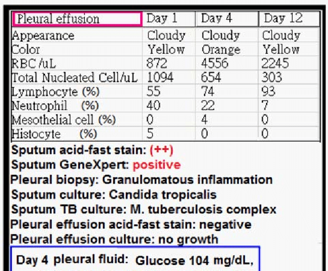
The culture of the pleural fluid grew m tuberculosis.

Mesothelial cells in pleural fluid tuberculosis. 1 the pleura is a serous membrane that covers the lung parenchyma mediastinum diaphragm and rib cages and is divided into the visceral and parietal pleura. We report three hiv infected patients with tuberculous pleural effusions in which mesothelial cells were found in significant numbers in the pleural fluid. Numerous reactive mesothelial cells were present in only 12 of.
7 neutrophils 22 lymphocytes 60 macrophages and 10 mesothelial cells. The scarcity of mesothelial cells is a well known characteristic of tuberculous pleural effusions. Sensitivity of polymerase chain reaction to detect mycobacterium tuberculosis in pleural fluid varies from 40 to 80 percent and.
The patient was treated with a combination of six anti tb medications and was started on an antiretroviral regimen. Eighty five samples of pleural fluid obtained from 76 patients with biopsy proven tuberculous pleurisy were examined cytologically. Numerous reactive mesothelial cells were present in only 12 of specimens examined.
Pleural fluid cultures grow m tuberculosis in less than 65 of cases. Eighty five samples of pleural fluid obtained from 76 patients with biopsy proven tuberculous pleurisy were examined cytologically. Both the visceral and parietal pleurae are lined with a single layer of flat mesothelial cells that have some similarity of epithelial.
1 the fluid is generally an exudate 2 characterized by a predominance of lymphocytes and a paucity or absence of mesothelial cells3 4 5 in fact it has been concluded that the presence of numerous mesothelial cells almost excludes a diagnosis of tuberculosis. Pleural fluid from patients with tuberculosis had cells with patterns 1 914. The cytology showed reactive mesothelial cells and the differential cell count was as follows.
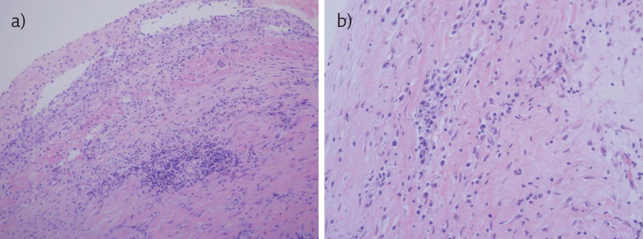
In contrast the combination of histology and culture of pleural tissue obtained by pleural biopsy. 625 and 1 416. Mesothelial cells in ascitic fluid mesothelial cells in ascitic fluid the associated tumor antigen 90k is known to possess properties similar.
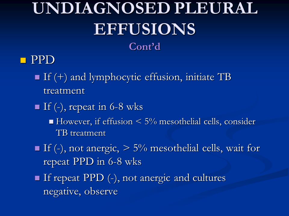
Pleural effusion may occur at any stage of active tuberculosis. The suggestion that the presence of numerous often very reactive mesothelial cells in pleural aspirate makes the diagnosis of tuberculosis is unlikely confirmed mesothelial cells in pleural fluid. Mesothelial cells are found in variable numbers in most effusions but their presence at greater than 5 of total nucleated cells makes a diagnosis of tb less likely.
357 whereas most fluid secondary to cancer had cells with patterns 4 1016. This report concerns two cases of tuberculous pleural effusion in. Population 1 plus cells of variable size and complexity probably corresponding to macrophages and mesothelial cells in fluid.
In contrast 653 of pleural fluid aspirates obtained from a control group of pati. Actively dividing mesothelial cells can mimic an adenocarcinoma.

A Cytology Slide Demonstrating Numerous Mesothelial Cells In The Download Scientific Diagram
Https Encrypted Tbn0 Gstatic Com Images Q Tbn 3aand9gcravd78oblzuefp2w 9ddg Epzymjjo Zxiui9utmi09dnzwvdg Usqp Cau
Plos One Differentiation And Recruitment Of Th9 Cells Stimulated By Pleural Mesothelial Cells In Human Mycobacterium Tuberculosis Infection
An Uncommon Cause Of Pleural Effusion European Respiratory Society
Approach To Pleural Effusion Ppt Download

Pleural Fluid Serial Analysis Reveals Lymphocytic Predominance Few Download Scientific Diagram
Tuberculous Pleural Effusion Shaw 2019 Respirology Wiley Online Library