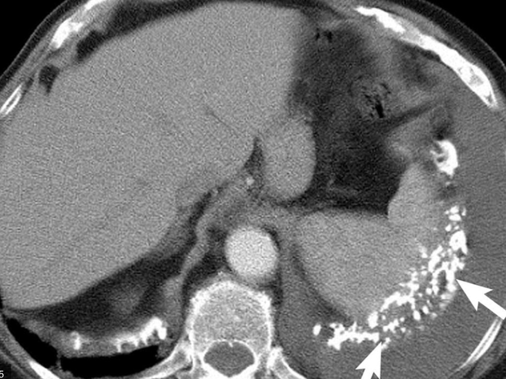
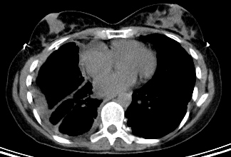
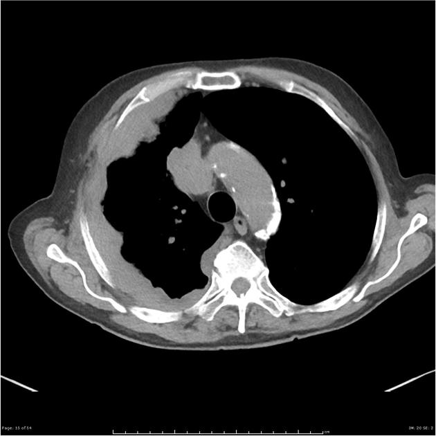
The connecticut mesothelioma victims centers services are available to people with mesothelioma or asbestos exposure lung cancer in new london groton bridgeport new haven hartford stamford. Ct demonstrates similar nonspecific findings.
Https Encrypted Tbn0 Gstatic Com Images Q Tbn 3aand9gcqm2wnxdnjwxjiarbr9ietjmckxbf5eolhcrkbcawdyim9zjp0n Usqp Cau
Crossref medline google scholar.

Mesothelioma ct. Ct scan of a patient with mesothelioma coronal section the section follows the plane that divides the body in a front and a back half. Ct manifestations in 50 cases. Ajr am j roentgenol 1990.
1 right lung 2 spine 3 left lung 4 ribs 5 descending part of the aorta 6 spleen 7 left kidney 8. Additional evidence of asbestos exposure such as calcified or noncalcified pleural plaques may be evident. Mesothelioma also known as malignant mesothelioma is an aggressive malignant tumor of the mesothelium.
This scan also can show if it has spread to lymph nodes or other organs. Diagnostic ct scan for mesothelioma. However with a history of significant asbestos exposure a diagnosis of mesothelioma can be suggested.
Mesothelioma is a malignant neoplasm originating from pleural or peritoneal surfaces. Ct stands for computerized tomography. In addition to the previously mentioned findings ct can also be useful in demonstrating invasion of the tumor into the mediastinum chest wall diaphragm and pericardium which aids in disease.
Ct scans can help physicians to determine the stage of your tumor. When a person goes to the doctor with early signs of malignant pleural mesothelioma a ct scan of the chest is often the next step in making a diagnosis. 15 kawashima a libshitz hi.
Appearances of asbestosis vary with the duration and severity of the condition. A new technique called ct perfusion can show if cancer cells are spreading in the bloodstream. Given the presence of the mesothelium in different parts of the body mesothelioma can arise in various locations 17.
Ct scans create a 3 dimensional image of internal organs making it easier to spot mesothelioma tumors. Pleural mesothelioma 90 covered in this article. The mesothelioma is indicated by yellow arrows the central pleural effusion fluid collection is marked with a yellow star.
More is better a new report out of the uk suggests that a thorough diagnostic ct scan for pleural mesothelioma should include images of the abdomen and pelvis as well as the chest. This condition is usually associated with occupational exposure to asbestoswagner et al connected asbestos to mesothelioma in a classic 1960 study of 33 patients with mesothelioma who were exposed to asbestos in a mining area in south africas north western cape province. How a diagnostic ct scan works.
But a research team led by the university of bristol says a diagnostic ct scan that shows only the chest might miss mesothelioma tumors in other areas. Still many doctors say the ct scan is the best for the chest and abdomen which are where mesothelioma forms. Most tumors arise from the pleura and so this article will focus on pleural mesothelioma.
Chest radiograph may show irregular opacities with a fine reticular pattern. Ct scanning is an.

Mesothelioma What Is The Mesothelioma Mesothelioma Dugli

Mesothelioma Symptoms Diagnosis And Treatment Bmj Best Practice Us

Malignant Pleural Mesothelioma Evaluation With Ct Mr Imaging And Pet Radiographics
Https Pubs Rsna Org Doi Pdf 10 1148 Rg 241035058

Pdf Oesphageal Stenting For Palliation Of Malignant Mesothelioma

Chest Ct Presenting The Mesothelioma Download Scientific Diagram
