However with a history of significant asbestos exposure a diagnosis of mesothelioma can be suggested. Less commonly the lining of the abdomen and rarely the sac surrounding the heart or the sac surrounding the testis may be affected.

Diagnostic Imaging And Workup Of Malignant Pleural Mesothelioma
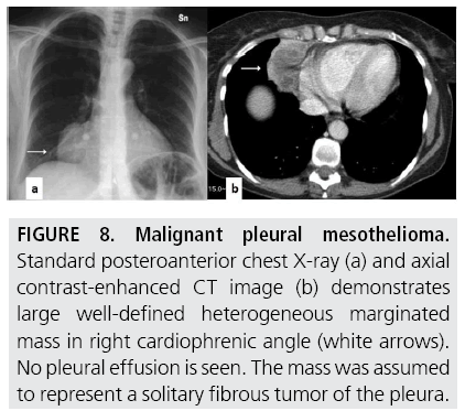
Given the presence of the mesothelium in different parts of the body mesothelioma can arise in various locations 17.

Mesothelioma ct findings. This scan also can show if it has spread to lymph nodes or other organs. Pleural thickening was found in 46 92 of the 50 patients thickening of the pleural surfaces of the interlobar fissures in 43 86 pleural calcifications in 10 20 and pleural effusions in 37 74. Pretreatment ct findings from 50 patients with malignant pleural mesothelioma are illustrated.
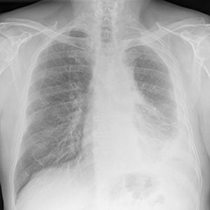
Generally abnormal chest x rays are followed by a more advanced imaging test such as a ct scan. Signs and symptoms of mesothelioma may. Ct is the primary imaging modality used for the evaluation of mpm.
Ct findings can delineate the optimal site for biopsy while providing a tremendous amount of anatomic information about the stage of the disease 16 20. The most common area affected is the lining of the lungs and chest wall. Yendamuri listed the following as common radiological findings in mesothelioma imaging exams.
Growth typically leads to tumoral encasement of the lung with a rindlike appearance. Mesothelioma is a type of cancer that develops from the thin layer of tissue that covers many of the internal organs known as the mesothelium. Pleural mesothelioma 90 covered in this article.
A new technique called ct perfusion can show if cancer cells are spreading in the bloodstream. Many of the tests are non invasive and they help physicians to determine the cause of certain symptoms. Ct scan findings are similar to those of plain films but are seen better and in more detail.
Mesothelioma also known as malignant mesothelioma is an aggressive malignant tumor of the mesothelium. Mesothelioma x ray findings ct scan and images if it is suspected that someone has mesothelioma or any other form of cancer a number of tests will be ordered. Mesothelioma awareness and understanding its signs is especially important for helping to diagnose mesothelioma at an early stage.
Most tumors arise from the pleura and so this article will focus on pleural mesothelioma. Nevertheless ct remains to be the dominant modality for assessing patients with mesothelioma including evaluation of treatment response. Ct demonstrates similar nonspecific findings.
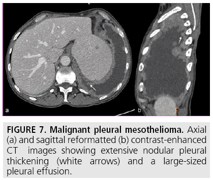
Key ct findings that suggest mpm include unilateral pleural effusion fig 1 nodular pleural thickening figs 2 4 and interlobar fissure thickening fig 5. Still many doctors say the ct scan is the best for the chest and abdomen which are where mesothelioma forms. In addition to the previously mentioned findings ct can also be useful in demonstrating invasion of the tumor into the mediastinum chest wall diaphragm and pericardium which aids in disease.
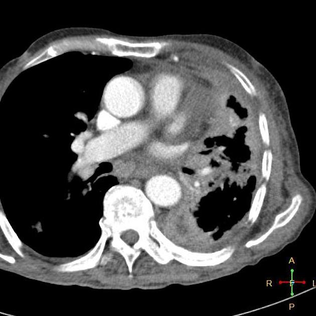
Here we show the ct scan of the chest from the same patient diagnosed with advanced mesothelioma. Ct scans can help physicians to determine the stage of your tumor. Furthermore pleural thickening and effusion can be distinguished with ct scanning.

Diagnostic Imaging And Workup Of Malignant Pleural Mesothelioma

Mesothelioma Diagnosis Understand The Diagnostic Process
Http Pdf Posterng Netkey At Download Index Php Module Get Pdf By Id Poster Id 106714
Http Bmb Oxfordjournals Org Content 93 1 105 Full Pdf 2bhtml

31 30 Axial Ct Scan Of A Patient With A Left Sided Malignant Download Scientific Diagram

Metastatic Biphasic Pleural Mesothelioma Presenting With Cauda Equina Syndrome Sciencedirect

The Role Of Imaging In Malignant Pleural Mesothelioma An Update After The 2018 Bts Guidelines Clinical Radiology
