Signs and symptoms of mesothelioma may. Mesothelioma is a cancer affecting the mesothelial cells which cover most internal organs.

Transmission Electron Microscopy Of A Mesothelioma Cell Prepared From Download Scientific Diagram
In 2018 there were 726 deaths caused by mesothelioma in australia.

Mesothelioma microscopy. The most common area affected is the lining of the lungs and chest wall. Less commonly the lining of the abdomen and rarely the sac surrounding the heart or the sac surrounding the testis may be affected. In 2016 774 people were diagnosed with mesothelioma in australia.
If you believe you may have mesothelioma you should get input from a specialist. This is according to an article published in the journal of thoracic disease titled the use of electron microscopy for diagnosis of malignant pleural mesothelioma electron microscopy which was first developed in the 1930s uses a beam of accelerated electrons to magnify an image of a structure to a much higher degree of magnification. An electron microscope is a powerful tool which allows for superior resolution of the specimen with the ability to magnify an object up to two million times.
In patients with cancer involving the lungs liquid often accumulates outside the lungs a feature called pleural effusion. Dominguez malagon h cano valdez am gonzalez carrillo c et al. What is malignant mesothelioma electron microscopy.
Electron microscopy is already in use to aid the diagnosis of mesothelioma when light microscopy analysis is inconclusive. They will utilize pathologists who have experience detecting this cancer and performing mesothelioma histology. Mesothelioma is also rare and many pathologists do not have experience studying and identifying mesothelioma cells under a microscope.
There are two main types of mesothelioma. Electron microscopy is a diagnostic tool using a type of microscopy which utilizes electrons to create an image. Mesothelioma is a type of cancer that develops from the thin layer of tissue that covers many of the internal organs known as the mesothelium.
But the tissue samples needed for this type of analysis need to be of high quality. Diagnostic efficacy of electron microscopy and pleural effusion cytology for the distinction of pleural mesothelioma and lung adenocarcinoma.

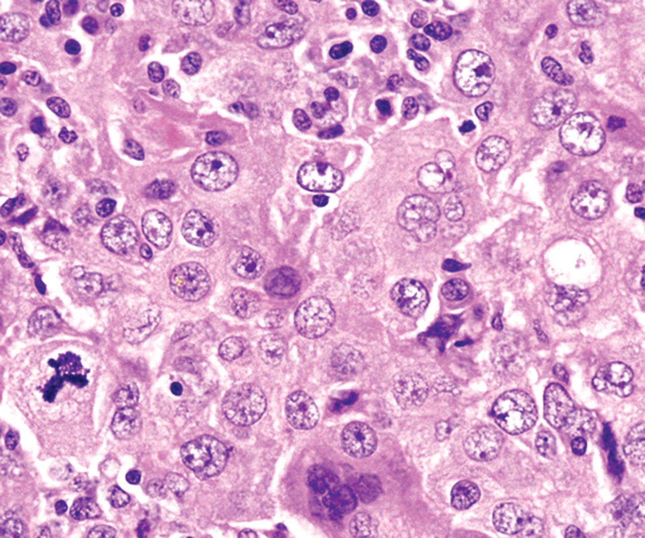
Malignant And Borderline Mesothelial Tumors Of The Pleura Thoracic Key

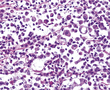
A Light Microscopy Of Mesothelioma In Situ Cells Showing A Download Scientific Diagram

Mesothelioma Scientific Clues For Prevention Diagnosis And Therapy Carbone 2019 Ca A Cancer Journal For Clinicians Wiley Online Library

Mesothelioma Oncology Medbullets Step 2 3

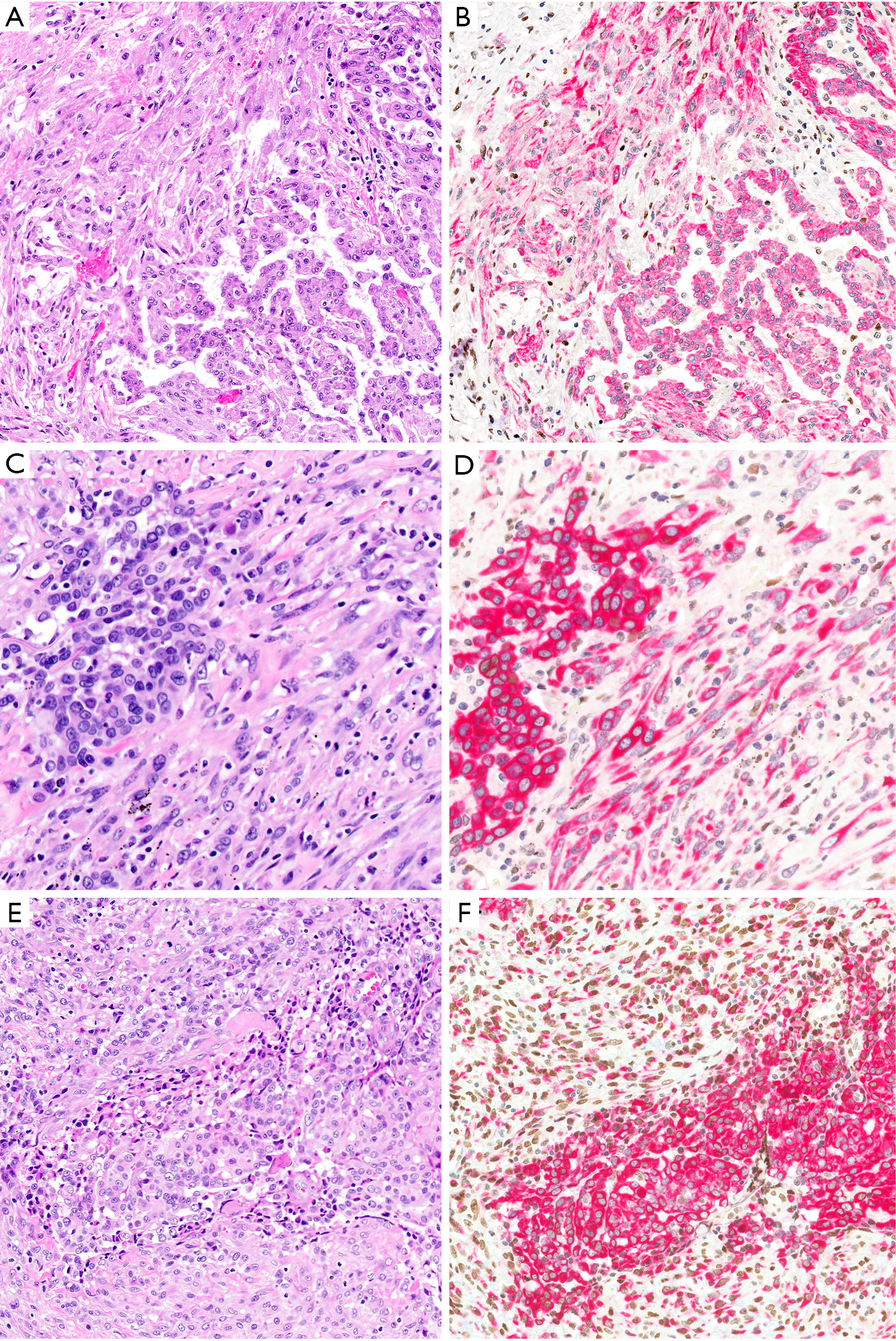
Application Of Immunohistochemistry In Diagnosis And Management Of Malignant Mesothelioma Chapel Translational Lung Cancer Research

Challenges And Controversies In The Diagnosis Of Malignant Mesothelioma Part 2 Malignant Mesothelioma Subtypes Pleural Synovial Sarcoma Molecular And Prognostic Aspects Of Mesothelioma Bap1 Aquaporin 1 And Microrna Journal Of Clinical Pathology
Https Link Springer Com Content Pdf 10 1007 2f0 387 28274 2 33 Pdf
