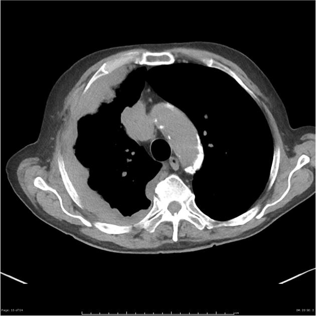
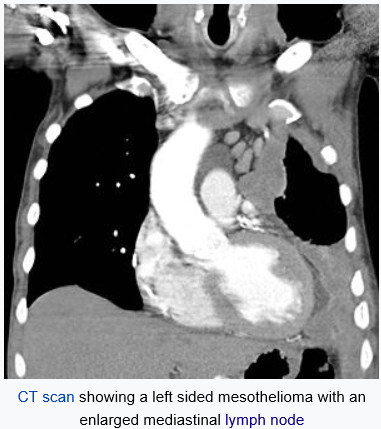
This upper ct scan slice reveals the calcified pleural plaques along the diaphragmatic surface that are associated with asbestos exposure. Ct scan of a patient with mesothelioma coronal section the section follows the plane that divides the body in a front and a back half.

Primary Peritoneal Mesothelioma Radiology Case Radiopaedia Org
Computed tomography ct scan in a male veterans administration patient with a history of asbestos exposure and an enlarging abdominal girth.

Mesothelioma on ct. Additional evidence of asbestos exposure such as calcified or noncalcified pleural plaques may be evident. Our team is working and offering consultations via phone e mail and video conferencing. Learn more about this process and other diagnosis issues from the illinois mesothelioma attorneys at shrader associates llp.
Chest radiograph may show irregular opacities with a fine reticular pattern. Typically patients with pleural mesothelioma receive a ct of the chest with contrast dye. The journal of thoracic disease indicates that not only does mesothelioma show up on a ct scan but it is the preferred diagnostic tool of choice for advanced stage mesothelioma cases.
J thorac cardiovasc surg 1988. Ct scans typically take 10 to 15 minutes to perform. Given the presence of the mesothelium in different parts of the body mesothelioma can arise in various locations 17.
Pleural mesothelioma 90 covered in this article. Ct mr and fdg pet. These imaging tests are frequently used when making a mesothelioma diagnosis because they may help pinpoint the location of cancer in the body.
1 right lung 2 spine 3 left lung 4 ribs 5 descending part of the aorta 6 spleen 7 left kidney 8. A new technique called ct perfusion can show if cancer cells are spreading in the bloodstream. Most tumors arise from the pleura and so this article will focus on pleural mesothelioma.
Ct scans are preferred for staging tumors and are vital for patients with malignant pleural mesothelioma yendamuri explained. Still many doctors say the ct scan is the best for the chest and abdomen which are where mesothelioma forms. Ct scans are often used to help diagnose mesothelioma.
Mesothelioma could show up on a ct scan or computed tomography scan especially if the ct scan is intentionally trying to look for it. This scan also can show if it has spread to lymph nodes or other organs. Ct scans can help physicians to determine the stage of your tumor.
Mesothelioma also known as malignant mesothelioma is an aggressive malignant tumor of the mesothelium. The mesothelioma is indicated by yellow arrows the central pleural effusion fluid collection is marked with a yellow star. Ascites is seen lateral to the liver.
The 3 d images ct scans provide a far more detailed view and offer more than 90 percent detection sensitivity. The role of computed tomography scanning in the initial assessment and the follow up of malignant pleural mesothelioma. Imaging of mediastinal lymph nodes.
18 rusch vw godwin jd shuman wp. Appearances of asbestosis vary with the duration and severity of the condition.

Primary Peritoneal Mesothelioma Radiology Case Radiopaedia Org

Mesothelioma Image Radiopaedia Org

Medpix Case Malignant Mesothelioma Epithelioid Type
Https Encrypted Tbn0 Gstatic Com Images Q Tbn 3aand9gcqwc6oe7ttcb Hourhci6giifatc6iektdkoon6yewe6pzkhm69 Usqp Cau

Localized Malignant Pleural Sarcomatoid Mesothelioma Misdiagnosed As Benign Localized Fibrous Tumor Kim Journal Of Thoracic Disease

Malignant Pleural Mesothelioma Evaluation With Ct Mr Imaging And Pet Radiographics

