Involving ipsilateral parietal pleura inc. Pleural mesothelioma 90 covered in this article.

Diagnostic Imaging And Workup Of Malignant Pleural Mesothelioma
Primary tumor cannot be assessed.

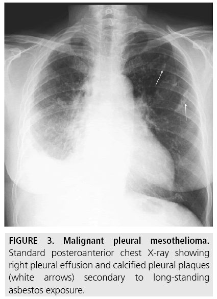
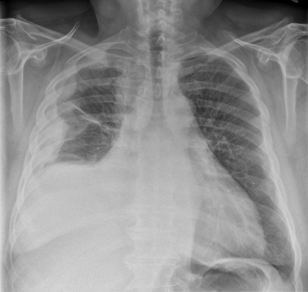
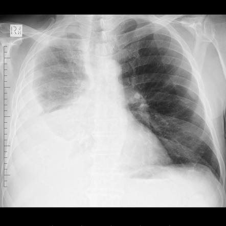
Mesothelioma pleural effusion radiology. Treatment involves draining the fluid but if it continues to accumulate more permanent procedures may be required. Malignant pleural mesothelioma mpm is an uncommon tumor of the pleura that can present a diagnostic challengecurrently the definitive diagnosis of mpm always is based on the results obtained from an adequate biopsy sample in the context of appropriate clinical radiologic and surgical findings. While a pleural effusion may be a symptom of pleural mesothelioma itself the condition can also cause its own symptoms like breathlessness.
Mesothelioma also known as malignant mesothelioma is an aggressive malignant tumor of the mesothelium. No evidence of primary tumor. For pleural mesothelioma patients pleural effusions develop in the majority of cases especially among patients with late stage disease.
Given the presence of the mesothelium in different parts of the body mesothelioma can arise in various locations 17. Most tumors arise from the pleura and so this article will focus on pleural mesothelioma. Pleural effusion is a buildup of fluid in the space around the lungs.
Involving any of the ipsilateral pleural surfaces with at least one of the. This case report is interesting and important because chest x ray and chest ct have characteristics of pleural abnormalities that can be used as guidelines for the diagnosis of mesothelioma. Mesothelioma also known as malignant mesothelioma is an aggressive malignant tumour of the mesothelium.
Pleural effusions are a common diagnosis in the united states and generally indicate a larger condition or disease. Pleural effusions are abnormal accumulations of fluid within the pleural space. Mesothelioma is a malignancy pleural mass characterized by pleural pleural effusionempyema.
Given the presence of the mesothelium in different parts of the body mesothelioma can arise in various locations 17. Pleural plaques confirming prior asbestos exposure and then a malignant effusion with pleural thickeni. Below is the eighth edition of the tnm staging system for malignant pleural mesothelioma which was published in 2018 1.
Most tumours arise from the pleura and so this article will focus on pleural mesothelioma. Pleural mesothelioma 90 covered in this article. Pleural effusion is a build up of fluid between the two layers of the pleura the thin tissue that surrounds the lungs and chest cavity.
Mediastinal and diaphragmatic pleura visceral pleura. Computed tomography ct has been used widely as the primary imaging modality. It is a typical complication of pleural mesothelioma and is uncomfortable or even painful.
This case has all the clinical and radiological features indicative of mesothelioma. Over time this fluid restricts the lungs and makes it hard to breathe. A prior profession in which asbestos exposure is common.
A new study says when it comes to pleural effusion in mesothelioma the two main solutions are pretty equal with one exception. Terminology pleural effusion is commonly used as. They may result from a variety of pathological processes which overwhelm the pleuras ability to reabsorb fluid.

Pleural Irregularities And Mediastinal Pleural Involvement In Early Stages Of Malignant Pleural Mesothelioma And Benign Asbestos Pleural Effusion European Journal Of Radiology

Mesothelioma Radiology Case Radiopaedia Org

Pleura Chest Wall And Diaphragm Chest Radiology The Essentials 2nd Edition

Pleural Effusions Mesothelioma

Figure 2 From Oral Presentation Of Malignant Mesothelioma Semantic Scholar


