
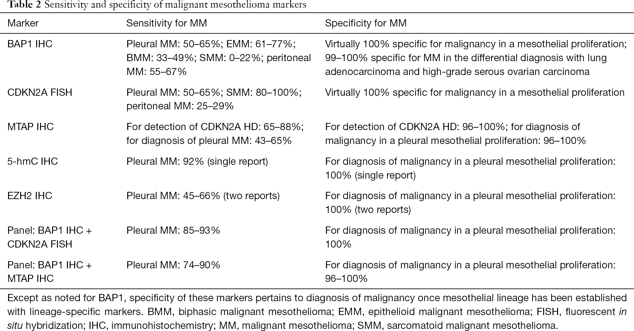
Several panel exists no agreed upon best panel. Cytology fna cytology effusions primary dx localized nodules vs.
Mesothelioma Vs Reactive Mesothelial Cells

There were no falsepositive diagnoses of mm in effusion specimens during this time period.
Mesothelioma vs adenocarcinoma cytology. Mesothelioma vs adenocarcinoma cytology. Adenocarcinoma is a subtype of non small cell lung cancer and it usually starts in the glands in the lungs. Usually two carcinoma markers two mesothelial markers.
Lioma and 925 of malignant mesothelioma versus 925 of adenocarcinoma. Mesothelioma specialists tend to have the best results when drawing cytology samples as they know exactly which parts of the body are most likely to collect mesothelioma cells. Conventional cytomorphologic assessment is the rst step to establish an accurate diagnosis in pleural effusions.
Most often doctors identify mesothelioma because of other problems the diseases cause. Patients who suspect a mesothelioma diagnosis should therefore seek out the help and expertise of a qualified mesothelioma specialist. Cea monoclonal and polyclonal.
Metastatic disease mesothelial cell lesions of pleura solitary fibrous tumor nodular pleural plaque adenomatoid tumor simple mesothelial cyst multicystic mesothelioma well differentiated papillary mesothelioma localized malignant mesothelioma. They each have different causes and prognoses and require very different treatment approaches. A history of asbestos exposure is a huge indicator for doctors to look for mesothelioma traits.
A mesothelioma diagnosis can be confused with lung cancer as they have similar sets of symptoms. Diagnosing mesothelioma vs adenocarcinoma. Mesothelioma and adenocarcinoma are both types of cancer but vary a great deal.
Most pathologists are reluctant to make a definitive diagnosis solely on fluid samples. Furthermore you may get more reliable information about the cell type from a histology report rather than just a cytology report. Of 217 cases circulated among all members of the uscanadian mesothelioma reference panel there was some disagreement about whether the process was benign or malignant in 22 of cases.
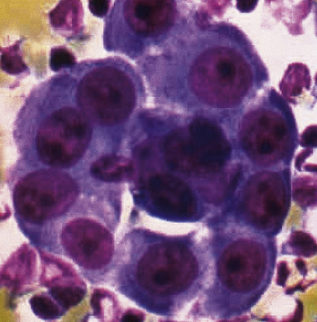
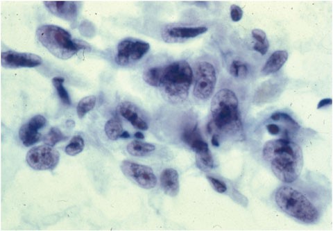
Several cytologic features have predictive value to seperate malignant mesothelioma from adenocarcinoma and reactive mesothelial proliferation. 1 frank invasion is regarded as the most. The distinction between reactive mesothelial hyperplasia mh and malignant mesothelioma mm may be very difficult based only on histologic and morphologic findings.
In addition 6 cases were designated atypical 2 were misclassified as positive for adenocarcinoma 1 was suspicious for mesothelioma and the remainder were classified as benign. Mesothelioma pathologists almost always request a tissue biopsy following a mesothelioma cytology report.
Pathologic Diagnosis And Classification Of Mesothelioma Springerlink

Flowchart Describing The Experimental Flow In Designing A Test With Download Scientific Diagram
Application Of Immunohistochemistry In Diagnosis And Management Of Malignant Mesothelioma Chapel Translational Lung Cancer Research
Https Www Hkiap Org Wp Content Uploads Lecture Notes 2019 20special 20cytology 20workshop Mesothelioma 20how 20far Pdf

Diagnostic Value Of Immunopathology In Malignant Pleural Tumors Farag Ts Farag As Oreiby Ha Egypt J Chest Dis Tuberc
Malignant And Borderline Mesothelial Tumors Of The Pleura Thoracic Key

D3i71xaburhd42 Cloudfront Net E348512bb7d0aa271