Most tumors arise from the pleura and so this article will focus on pleural mesothelioma. Many of the tests are non invasive and they help physicians to determine the cause of certain symptoms.

Malignant Mesothelioma Imaging Overview Radiography Computed Tomography
This is the most common type of mesothelioma accounting for nearly 70 percent of cases.

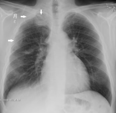
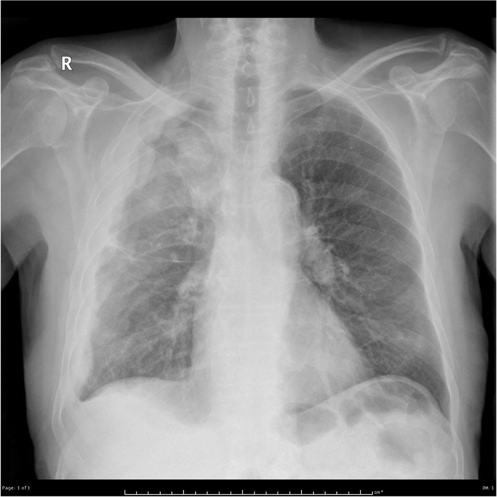
Mesothelioma x ray images. For people who may have mesothelioma an x ray helps doctors determine if there is an anomaly in their lungs. One of the biggest drawbacks of an x ray is that it can only produce a flat two dimensional image. Mesothelioma also known as malignant mesothelioma is an aggressive malignant tumor of the mesothelium.
The 3d image obtained from a ct scan provides a far more detailed view of a persons internal anatomy than a simple two dimensional x ray image. When an x ray is taken electromagnetic radiation is sent through the body with a photographic film on the other. Mesothelioma x ray findings ct scan and images if it is suspected that someone has mesothelioma or any other form of cancer a number of tests will be ordered.
X rays are used to examine all parts of the body including the chest and abdomen. This scan is limited but may be able to detect damage or abnormalities in the body. The most basic imaging scan is an x ray.
An x ray is a test that uses small doses of radiation to take pictures of the inside of your body. Given the presence of the mesothelium in different parts of the body mesothelioma can arise in various locations 17. An x ray uses electromagnetic beams to collect two dimensional images of the inside of your body.
They are a good way to look at bones and can show changes caused by cancer or other medical conditions. This is usually the first imaging test youll undergo. Pleural mesothelioma 90 covered in this article.
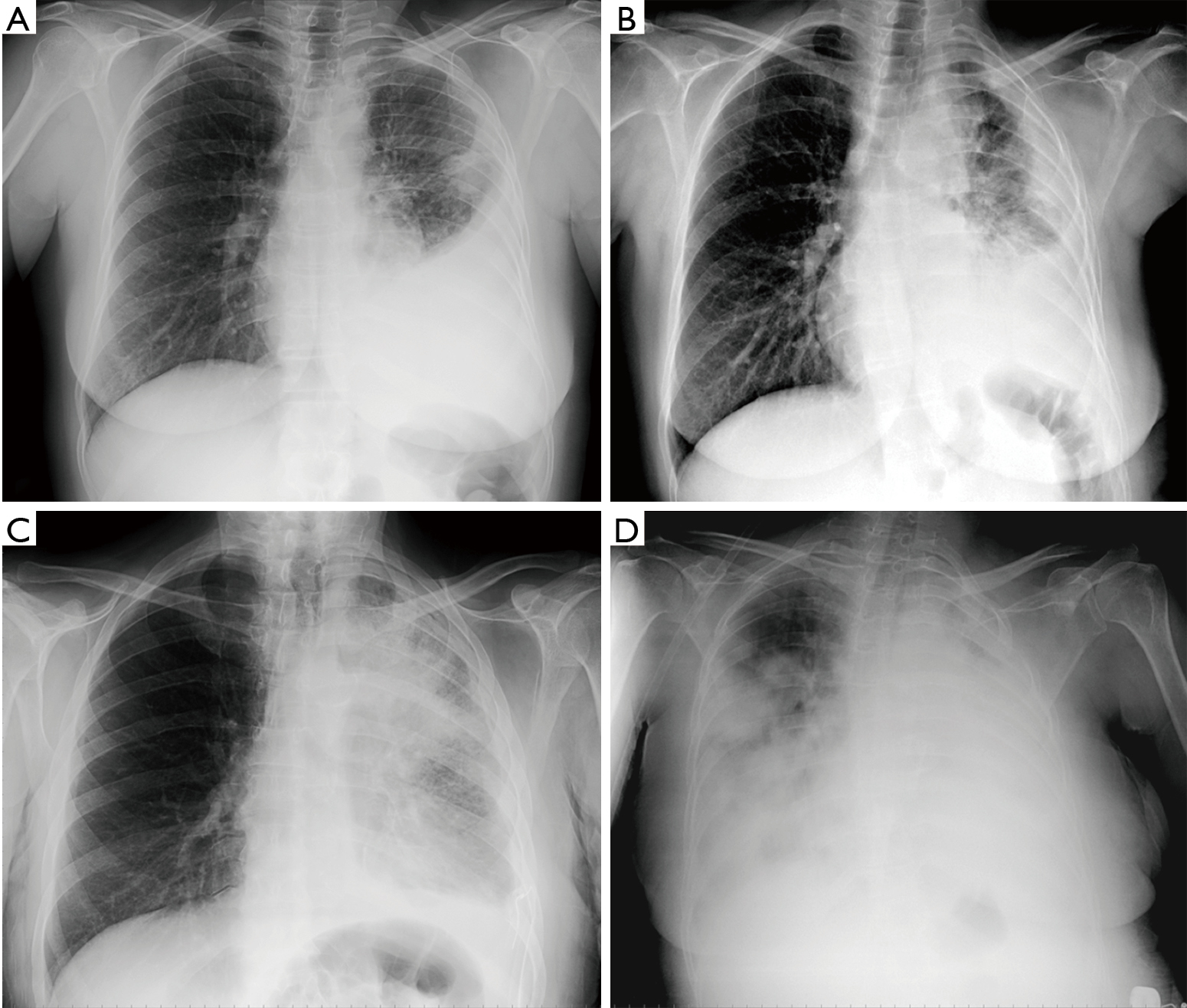
X rays are usually the first type of imaging used to investigate signs of mesothelioma and other diseases that affect the lungs or heart. X rays also indicate fluid build up another type of asbestos related condition which may indicate mesothelioma. Imaging of this type of the disease will depict the greater abdominal region as it attacks the lining of.
There are five primary types of imaging tests used during a mesothelioma diagnosis including x rays mri scans ct scans pet scans and ultrasounds. Ct scans are more than 90 sensitive for detecting malignant pleural mesothelioma. An x ray uses high energy electromagnetic radiation to image dense tissue in the body.
Imaging scans are non invasive and act as a stepping stone in the diagnostic process for mesothelioma patients. Using an x ray image the doctor can see if the pleura around the lungs has thickened an indication of cancer. X ray images of this kind of mesothelioma will depict the lungs as it affects the lining of the lungs.
Those who are at higher risk of developed asbestos cancer such as factory shipbuilding or construction workers may get a chest x ray as an initial screening tool. An x ray provides a flat 2d image of bones and soft tissue.

Clinical Diagnosis Of Malignant Pleural Mesothelioma Bianco Journal Of Thoracic Disease

Action Mesothelioma Day 2018 What Is Mesothelioma Oliver Co Solicitors Cheshire

Mesothelioma Chest X Ray On The Left Lung Mesothelioma

What To Expect When You Have Mesothelioma Pleural Effusion

Multimodality Imaging For Characterization Classification And Staging Of Malignant Pleural Mesothelioma Radiographics
Https Encrypted Tbn0 Gstatic Com Images Q Tbn 3aand9gct9qsrwofmw6gofhgqizvehuhysmpo Yjuxibedakrpolzogxig Usqp Cau

