There are certain cells that line the pleura the thin double layered lining which covers the lungs chest wall and diaphragm which are known as mesothelial cellsother than the pleura mesothelial cells also form a lining around the heart pericardium and the internal surface of the abdomen peritoneum. Careful measurements in rabbits and dogs have yielded volumes of pleural fluid of 01 to 03 mlkg and a similar volume is thought to be present in normal humans normal pleural fluid at least in laboratory animals such as rabbits and dogs 3 4 contains a significant number.

Cytology Of Pleural And Peritoneal Lesions Chapter 5 Practical Pathology Of Serous Membranes
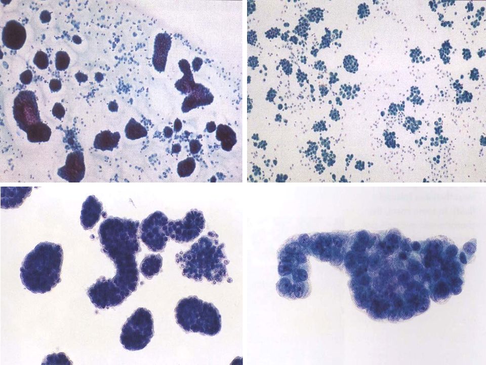
Many reactive mesothelial cells were present in only 12 of samples analyzed.
Normal mesothelial cells in pleural fluid. No tuberculous effusions had more than 1 mesothelial cells while most other effusions contained at least 5 mesothelial cells. Ada is an enzyme that plays an important role in lymphoid cell differentiation. Pleural fluid cytological studies showed malignant cells in 33 of 43 patients with effusions due to tumor.
It deals with pericardial fluid peritoneal fluid and pleural fluid. It is not uncommon for macrophages to be mixed in a ball reactive mesothelial cells. The mesothelium is composed of an extensive monolayer of specialized cells mesothelial cells that line the bodys serous cavities and internal organs.
In case of effusions due to pneumonic lesion a pleural fluid ph of less than 71 72 indicates the need for urgent drainage of the effusion while a pleural fluid ph of more than 73 suggests. A pleural fluid ph of less than 730 with a normal arterial blood ph level is caused by the same diagnoses as listed above for low pleural fluid glucose. A pleural fluid ada level greater than 40 u per l 667 nkat per l has a sensitivity of 90 to 100 percent and a.
In normal humans a small amount of pleural fluid is present 1 2the exact volume of this fluid is unknown. Mesothelial cytopathology is a large part of cytopathology. The article deals with cytopathology specimens from spaces lined with mesothelium ie.
Of 31 exudative effusions with a lymphocytic predominance 30 were due either to tuberculosis or neoplasm. This is known as pleural effusion. A patient may develop shortness of breath and vague chest pains while the fluid experiences a buildup in the chest cavity these are two of the most common.
The fluid can accumulate quickly if the mesothelial cells fail to function resulting in an unhealthy collection of fluids in the chest cavity. The main purpose of these cells is to produce a lubricating fluid that is released between layers providing a slippery non adhesive and protective surface to facilitate intracoelomic movement. An introduction to cytopathology is in the cytopathology article.
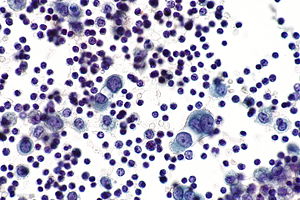
Markedly increased numbers of. Mesothelial cells in pleural fluid eighty five samples of mesothelial cells in pleural fluid from 76 patients with biopsy proven tuberculous pleurisy were examined cytologically. Mesothelial cells are found in variable numbers in most effusions but their presence at greater than 5 of total nucleated cells makes a diagnosis of tb less likely.
Mesothelial cells in pleural fluid.
Cytology Of Body Fluid Pleural Peritoneal Pericardial Ppt Video Online Download
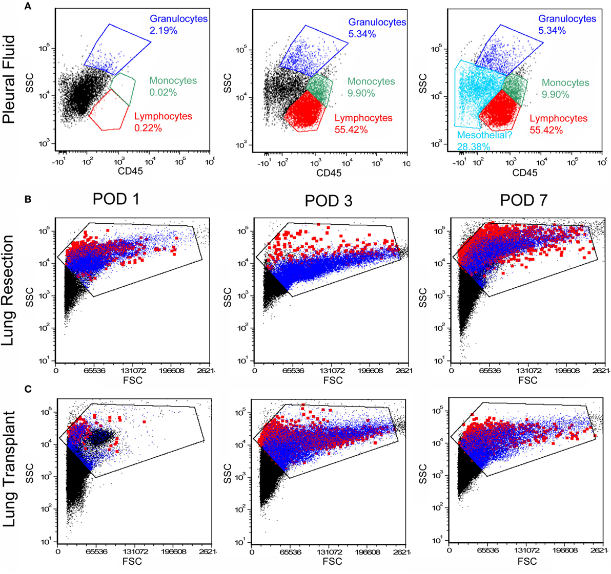
Frontiers Free Floating Mesothelial Cells In Pleural Fluid After Lung Surgery Medicine
Http Www Api Pt Com Reference Commentary 2015ascope Pdf
Https Encrypted Tbn0 Gstatic Com Images Q Tbn 3aand9gcqhd7waupdnvkltywmzp1zjlgtbh2fsowenirnocak8jd8 Jgve Usqp Cau

Claudin 4 Expression In Mesothelial Cells From Inflammatory Pleural Download Scientific Diagram

Pleural Pericardial And Peritoneal Fluids Basicmedical Key
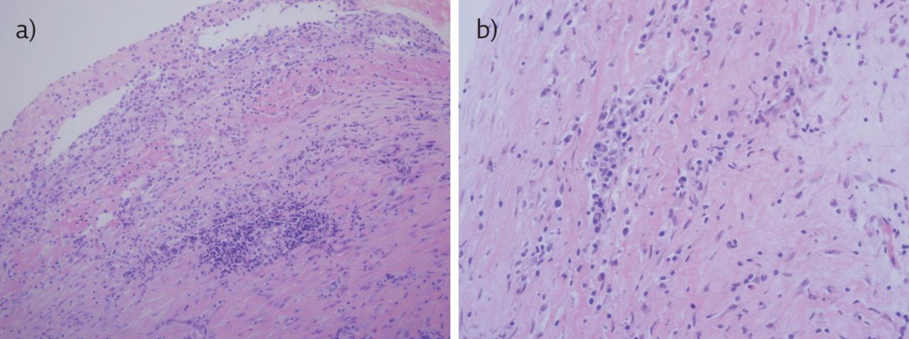
An Uncommon Cause Of Pleural Effusion European Respiratory Society