Small cell carcinoma isolated cells clusters and chains round or angular nuclei molding may show elongation and spindling nuclear molding. An introduction to cytopathology is in the cytopathology article.
Home
Ihc and pap cytology are more reliable pleural fluid from a 73 yo man.
Reactive mesothelial cells in pleural fluid cytology. Reactive mesothelial cells reactive mesothelial cells in pleural fluid reactive mesothelial cells are found when there is infection or inflammation present in a body cavity. Reactive mesothelial cells may sometimes be sampled in mediastinal fine needle aspirates and should not be mistaken as thymoma cells. It deals with pericardial fluid peritoneal fluid and pleural fluid.
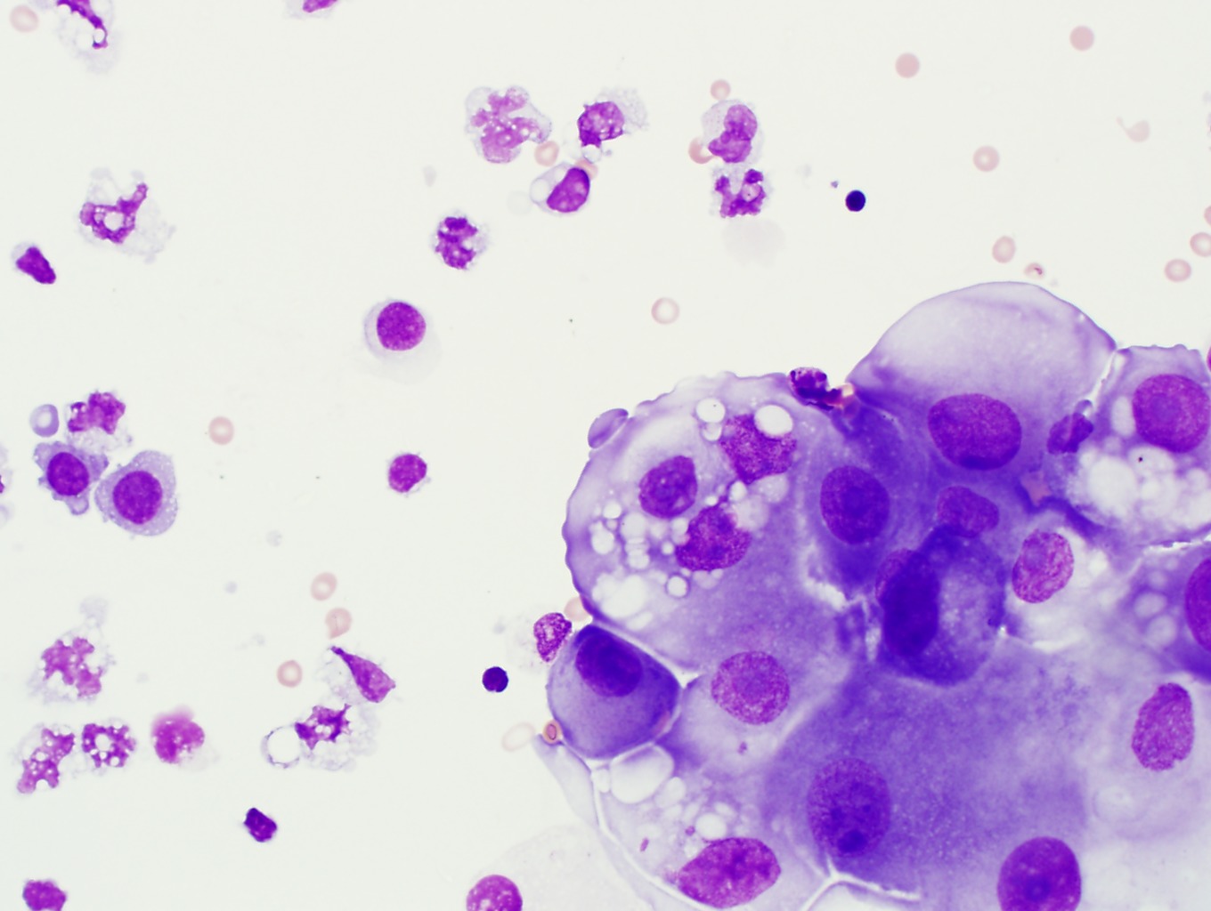
Fna lung pleural fluid. Although cytological examination helps in diagnosis of malignancy in serous effusion at times it is difficult to differentiate atypical reactive mesothelial cells from adenocarcinoma ac cells. Mesothelial cytopathology is a large part of cytopathology.
Neg in reactive mesothelial cells in 4 of cases. To resolve this problem various ancillary methods have been used. Further work up the tumor cells were also positive for wt1 calretinin figure 1c1 and andc2 c2 p53 and cytokeratin 56 and stained negatively for pax 8 berep4 desmin cea and ttf 1.
Actively dividing mesothelial cells can mimic an adenocarcinoma. Junko ueda takako iwata midori ono mutsuo takahashi comparison of three cytologic preparation methods and immunocytochemistries to distinguish adenocarcinoma cells from reactive mesothelial cells in serous effusion diagnostic cytopathology 101002dc20391 34 1 6 10 2005. Immunocytochemistry icc is one such commonly used technique in which various panel of antibodies has been tried.
20 of 119 of patients with malignancy involving the pleura but a negative pleural fluid cytology. They contain ovoid nuclei fine chromatin delicate nuclear membrane small nucleoli and a moderate. The reactive mesothelial proliferations form smaller uniform less complex groups compared to malignant mesothelial proliferations.
To evaluate the use of a panel of markers to differentiate adenocarcinoma and the reactiveinflammatory process in fluid cytology we stained 29 formalin fixed paraffin embedded cell blocks of effusion fluid from patients with metastatic adenocarcinoma and 24 cell blocks from patients with benign effusion with mucicarmine and antibodies to carcinoembryonic antigen cea b723 and calretinin. Benign mesothelial cells tend to arrange in monolayered sheets with little nuclear overlapping fig. The article deals with cytopathology specimens from spaces lined with mesothelium ie.
Neoplastic transformation of mesothelial cells results in malignant mesothelioma an aggressive tumor especially the pleura.
Effusion Cytomorphology Of Rhabdomyosarcoma A Rare Case Of Primary Mediastinal Rhabdomyosarcoma With Superior Vena Cava Obstruction And Bilateral Pleural Effusion
01 Presentation I Vs 8 55mb 3 28 08 Pps
Https Www Rcpath Org Asset Ed8cdd8d 8d04 4b82 Ad48d585e2f023be
Body Cavity Effusions Are Among The Most Commonly Received
![]()
Shutterstock Puzzlepix
Https Www Sciencedirect Com Science Article Pii S1877050913010909 Pdf Md5 Dcd46a4816e9d057d4912a8943bc10d6 Pid 1 S2 0 S1877050913010909 Main Pdf

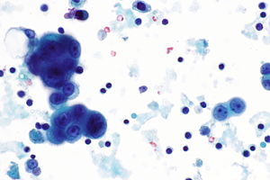
Mesothelioma In A Pleural Fluid Specimen Atypical Mesothelial Cells Download Scientific Diagram