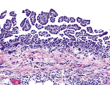
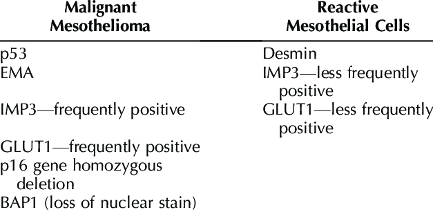
The differential diagnosis of epithelial type mesothelioma from adenocarcinoma and reactive mesothelial proliferation. Deletion of 9p21 and p16 are associated with inferior survival 345.

Common Immunohistochemical Stains Used To Differentiate Pulmonary Download Table
Focal macronucleoli are seen in reactive mesothelial cells.
Reactive mesothelial cells vs mesothelioma ihc. Lin md phd. 1 frank invasion is regarded as the most. P16 in mesothelioma and reactive mesothelium many mesotheliomas show deletion of the p16cdkn2a gene.
17 assessed ki 67 and other proliferation marker called repp86 and demonstrated that used in combination they are useful to discriminate between malignant mesothelioma and reactive mesothelial cells. 3 d clusters of cells strongly. Archival paraffin embedded cell blocks of pleural and peritoneal fluids from 52 patients with malignant mesothelioma mm and 64 patients with reactive mesothelial hyperplasia mh were retrieved.
Of 217 cases circulated among all members of the uscanadian mesothelioma reference panel there was some disagreement about whether the process was benign or malignant in 22 of cases. Large nc ratios may be seen in reactive mesothelial cells. Nc ratio may be normal in mesothelioma.
In situ hybridisation ish to demonstrate p16 deletions has been proposed as useful in differentiating between malignant mesothelioma and reactive mesothelial proliferations. The morphological evaluation of cytological specimens from body cavity fluids presents difficulties in the differential diagnosis between benign reactive mesothelial rm cells and adenocarcinoma ac or malignant mesothelioma mm. Reactive mesothelial proliferation 6 8 on morphology signs of malignancy include invasion for instance in the adipose tissue lung and skeletal muscle and the keratin really can be helpful to highlight the invasion of the neoplastic cells.
The aim of our study was to investigate whether a panel of five dif. Immunohistochemical detection of glut 1 can discriminate between reactive mesothelium and malignant mesothelioma. The use of immunohistochemistry to distinguish reactive mesothelial cells from malignant mesothelioma in cytologic effusions farnaz hasteh md 1.
Nuclear membrane irregularies rare. The distinction between reactive mesothelial hyperplasia mh and malignant mesothelioma mm may be very difficult based only on histologic and morphologic findings. Ihc stains included desmin epithelial membrane antigen ema glucose transport protein 1 glut 1 ki67 and p53.
Focal hyperchromasia is seen in reactive mesothelial cells. The distinction of benign from malignant mesothelial proliferations in cytologic specimens can be. 163299 305 26 kato y tsuta k seki k et al.
Mesotheliomas than in reactive mesothelial hyperplasia and better results obtained with mcm2 11 16.

Use Of Panel Of Markers In Serous Effusion To Distinguish Reactive Mesothelial Cells From Adenocarcinoma Subbarayan D Bhattacharya J Rani P Khuraijam B Jain S J Cytol

Malignant And Borderline Mesothelial Tumors Of The Pleura Thoracic Key

Utility Of Cell Block To Detect Malignancy In Fluid Cytology Adjunct Or Necessity Dey S Nag D Nandi A Bandyopadhyay R J Can Res Ther

Histology And Immunohistochemistry Of Lesions In Mextag Mice Following Download Scientific Diagram
Https Encrypted Tbn0 Gstatic Com Images Q Tbn 3aand9gcqvrwk0ao0 Cd6bc69apnaythotoaokap7n8inn B Xafzfzdo2 Usqp Cau
Https Www Surgpath Theclinics Com Article S1875 9181 10 00011 5 Pdf

Reliability Of P 16 Calretinin And Claudin 4 Immunocytochemistry In Diagnostic Verification Of Effusion Cytology
