Tag Archives: canine mesothelioma cytology

Canine Mesothelioma Cytology

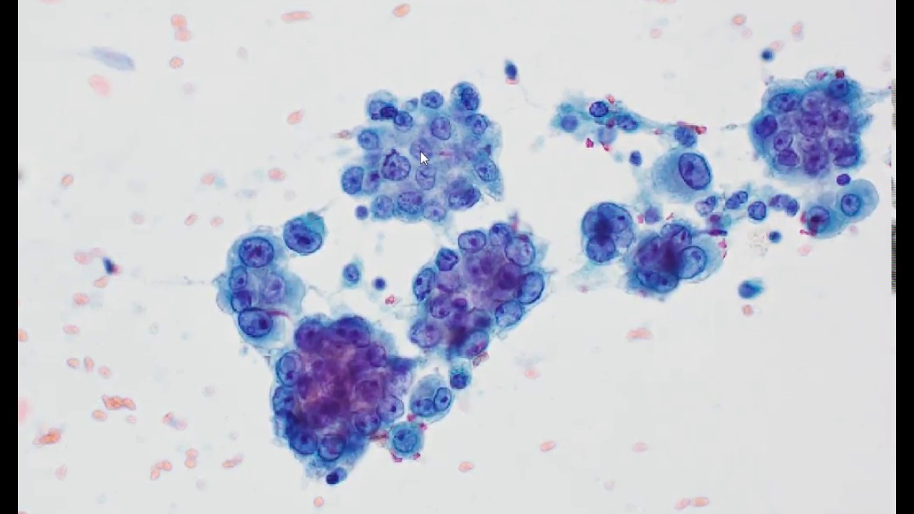
Mesothelioma cytology is the observe of most cancers fluid samples. It may be used to diagnose mesothelioma and calls for a minimally invasive fluid biopsy.


Effusion Cytology Clinician S Brief

Other checks together with a chest xray sputum cytology.

Canine mesothelioma cytology. Risk of mesothelioma has been associated with long term exposure to asbestos and pesticides. Mcdonough sp maclachlan nj tobias ah 1992 canine pericardial mesothelioma. Renal mass and localized renal cancer aua tenet.

If fluid has collected in the pericardial sac surgery to relieve the pressure will be required. 1 frank invasion is regarded as the most. The distinction between reactive mesothelial hyperplasia mh and malignant mesothelioma mm may be very difficult based only on histologic and morphologic findings.

Kobayashi y usuda h ochiai k et al 1994 malignant mesothelioma with metastases and mast cell leukaemia in a cat. Typically occurring in older dogs mesothelioma is more common in males than females. Of 217 cases circulated among all members of the uscanadian mesothelioma reference panel there was some disagreement about whether the process was benign or malignant in 22 of cases.

13 spontaneous mesothelioma has been reported in humans as well as in many species of animals including dogs cattle goats horses rats and hamsters 7 but is most common in cattle where it may be congenital. J comp pathol 111 4 453 458 pubmed. If your dog has an excess of fluid in any of its body cavities as a result of the mesothelioma such as in the chest or abdomen your veterinarian will need to hospitalize it for a short period of time in order to drain these cavities.

Moore a s 1992 chemotherapy for intrathoracic cancer in dogs and cats. Raflo c p nuernerger s p 1978 abdominal mesothelioma in a cat. Mesothelioma cytology pathology diagnosis by way of fluid.

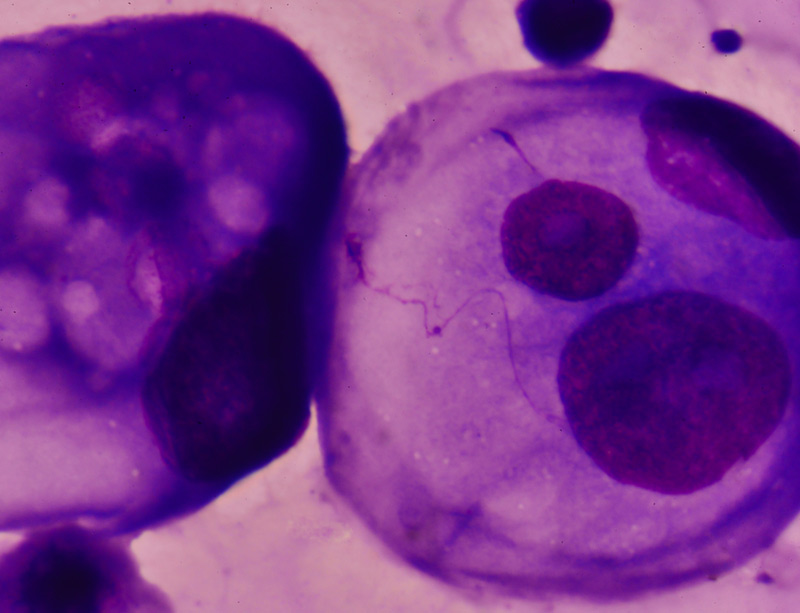
Mesothelioma is a rare neoplasm arising from the lining cells of the peritoneal pleural and pericardial cavities or the tunica vaginalis of the testis. The mesothelial cells in this fluid resemble those seen in many non neoplastic effusions and essentially lack cytologic criteria of malignancy despite this fluid being from a dog with documented mesothelioma based on tissue invasion in surgical biopsies of tissues. Vet pathol 29256 260 liu kx et al 1994 antigen expression in normal and neoplastic canine tissues defined by a monoclonal antibody generated against canine mesothelioma cells.

8 mesothelioma is associated. The main challenge in cytology analysis of mesothelioma is to distinguish it from reactive mesothelial cells considering that in both cases the cells present morphological atypia and increase of. Probl vet med 4 2 351 364 pubmed.

Atlas cytology canine peritoneal fluid canine peritoneal fluid. However it can occur in juveniles and females. Mesothelioma is a rare cancerous tumor in dogs.


Conference 7 2017 Case 2 20171018


Neoplastic Pleural Effusion And Intrathoracic Metastasis Of A Scapular Osteosarcoma In A Dog A Multidisciplinary Integrated Diagnostic Approach Mesquita 2017 Veterinary Clinical Pathology Wiley Online Library


Mesothelioma Dog


Soft Tissue Sarcomas With Epithelioid Morphology Chapter 5 Soft Tissue Sarcomas


A Descriptive Review Of Cardiac Tumours In Dogs And Cats Treggiari 2017 Veterinary And Comparative Oncology Wiley Online Library
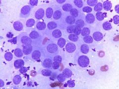


Bryan Clin Path Test 3 Winter 2nd Year Cytology Flashcards Cram Com
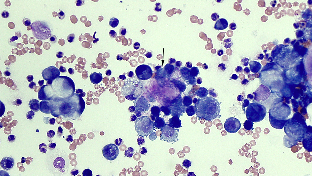


Cpd From Cloudy To Clear Effusions Made Easy Vet360

Mesothelioma Cytology

It deals with pericardial fluid peritoneal fluid and pleural fluid. Signs and symptoms of mesothelioma may.


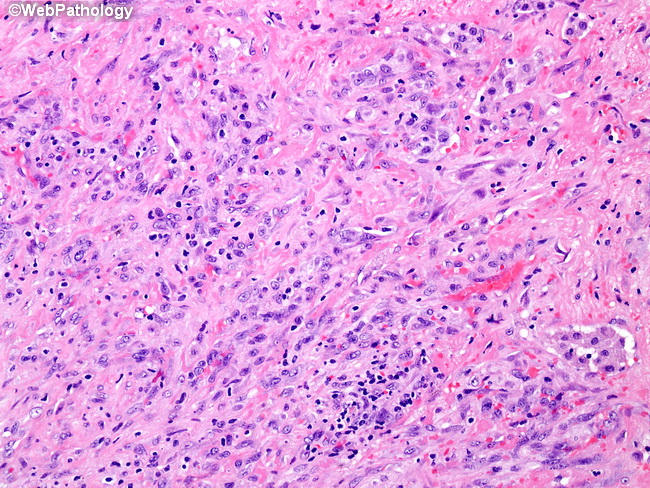
Webpathology Com A Collection Of Surgical Pathology Images

This fact is due to tissue samples being more reliable than fluid samples.

Mesothelioma cytology. It is a part of mesothelioma pathology which is the study of tissue or fluid to determine if this cancer exists. One study published on uptodate compared the accuracy of tissue biopsies and fluid samples. Mesothelioma cytology unlike imaging tests cytology is a pathological means to diagnosing malignant mesothelioma in patients consisting of a fluid biopsy of abnormalities previously discovered.

Mesothelioma cytology or cytopathology goes hand in hand with histology. However there is still a small risk of tumor cell seeding microscopic spreading of cancer due to cells moving during a biopsy during cytologic procedures. A look at how the cancer will advance.

Mesothelioma histology is more trusted than cytology for pathologically diagnosing this disease. By forgoing a tissue biopsy there is less risk of morbidity of the patient. Less commonly the lining of the abdomen and rarely the sac surrounding the heart or the sac surrounding the testis may be affected.

The article deals with cytopathology specimens from spaces lined with mesothelium ie. The most common area affected is the lining of the lungs and chest wall. While the histological determination will focus more on the cell type the cytology research will dive deeper into how these cells are forming interacting and spreading in the body.

Mesothelial cytopathology is a large part of cytopathology. Mesothelioma cytology or mesothelioma cytopathology is the study of cells for the presence of mesothelioma. Mesothelioma is a type of cancer that develops from the thin layer of tissue that covers many of the internal organs known as the mesothelium.

Cytology is often one of the first tests used in the diagnosis of mesothelioma after imaging tests reveal the possibility of cancer. Mesothelioma cytology is a useful tool that enables analysis without necessitating a tissue biopsy. Carlos wm bedrossian an update on pleuropulmonary cytopathology.

Once cytology is performed the cells from the sample are examined to arrive at a diagnosis. Cytology or fluid biopsy is a pathology specialty which studies disease by looking at the appearance and behavior of cells in bodily fluids. Mesothelioma cytology mesothelioma cytology.

Cytological diagnosis of mesothelioma and molecular cytology of lung cancer with an historical perspective diagnostic cytopathology 101002dc23298 43 7 513 526 2015.


Figure 2 A B Neoplastic Cells Arrows Intermixed With Reactive Mesothelial Cells Note The Eccentric Location Of The Nuclei Of Neoplastic Cells Kletki Krovi


Moving Toward An Earlier Diagnosis Of Mesothelioma


Cytological Profile Of A Case Of Malignant Mesothelioma Of Pleura With Supraclavicular Lymph Node Metastasis Abstract Europe Pmc


Https Onlinelibrary Wiley Com Doi Pdf 10 1002 Cncy 21342


Mesothelioma Dog


Https Encrypted Tbn0 Gstatic Com Images Q Tbn 3aand9gcsbgvpsjkfmxbyb2dtepoaktmrusji5uunvmadtoap8gksrx8cp Usqp Cau


The Value Of Brca 1 Associated Protein 1 Expression And Cyclin Dependent Kinase Inhibitor 2a Deletion To Distinguish Peritoneal Malignant Mesothelioma From Peritoneal Location Of Carcinoma In Effusion Cytology Specimens Althakfi 2020