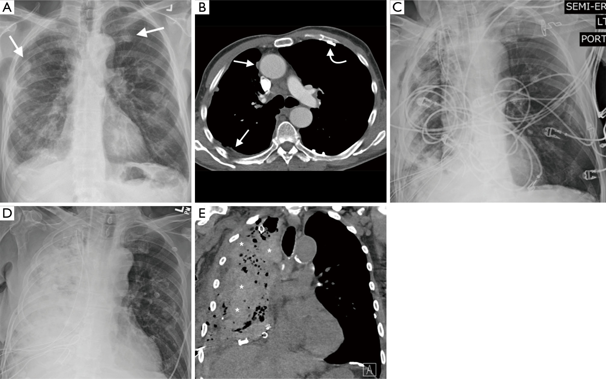
Tag Archives: ct scan mesothelioma radiology

Mesothelioma Ct Scan

The computer puts them together to make a 3 dimensional 3d image. Ct scan if doctors suspect mesothelioma in their patients they may recommend various diagnostic tools including medical imaging procedures.


Https Encrypted Tbn0 Gstatic Com Images Q Tbn 3aand9gcqm2wnxdnjwxjiarbr9ietjmckxbf5eolhcrkbcawdyim9zjp0n Usqp Cau

The computed tomography scan is an important tool in this process because it can provide highly detailed information about the disease type location and metastasis.

Mesothelioma ct scan. Ct scans can help physicians to determine the stage of your tumor. Ct scans are useful for diagnosing mesothelioma. A ct scan is a test that uses x rays and a computer to create detailed pictures of the inside of your body.

The image shows abnormal tissue which could be malignant tumors. Learn more about this process and other diagnosis issues from the illinois mesothelioma attorneys at shrader associates llp. This scan also can show if it has spread to lymph nodes or other organs.

It takes pictures from different angles. A new technique called ct perfusion can show if cancer cells are spreading in the bloodstream. Ct or cat stands for computed axial tomography.

Still many doctors say the ct scan is the best for the chest and abdomen which are where mesothelioma forms. You usually have a ct scan in the x ray radiology department as an. Ct scans also help doctors determine if treatment is working and the effect it has had on tumor number and size.

These scans also help stage cancer determining how much it has spread to other tissues. In short the best scientific evidence to date suggests that while ct scans are a useful tool in the work up of mesothelioma they cannot be relied upon to be fully accurate. Ultimately a biopsy is the only way to diagnose mesothelioma although the ct scan helps direct when and where to biopsy.

Ct scans are often used to help diagnose mesothelioma.
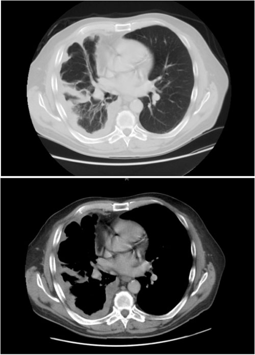


Diagnosis Of Malignant Pleural Mesothelioma Stanford Health Care


Pleural Effusion In Mesothelioma Ct Scan Stock Image C013 9667 Science Photo Library


The Role Of 18f Fdg Pet Ct Integrated Imaging In Distinguishing Malignant From Benign Pleural Effusion


Mesothelioma Lung Cancer Ct Scan Photograph By Du Cane Medical Imaging Ltd
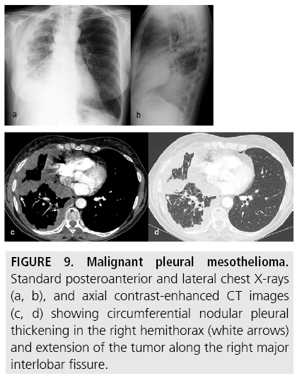


Diagnostic Imaging And Workup Of Malignant Pleural Mesothelioma


Pulmonary Radiologic Images


Metastatic Biphasic Pleural Mesothelioma Presenting With Cauda Equina Syndrome Sciencedirect
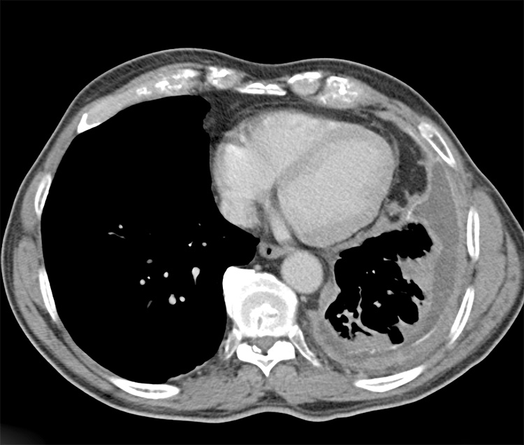
Mesothelioma Radiology Ct

Ct mr and fdg pet. 18 rusch vw godwin jd shuman wp.


Yazdir Kisa Rapor Short Report Doi 10 5578 Tt 10100 Tuberk Toraks 2016 64 3 253 255 Gelis Tarihi Received 09 08 2015 Kabul Edilis Tarihi Accepted 11 08 2015 Iki Farkli Primer T M Rden Kaynaklanan Ps Domezotelyoma Olgularinin G R Nt

Mesothelioma also known as malignant mesothelioma is an aggressive malignant tumor of the mesothelium.

Mesothelioma radiology ct. Asbestosis typically occurs 10 15 years following the commencement of exposure to asbestos and is dose related 3heavy asbestos exposure is predominantly encountered among men as most exposures are occupational in the setting of construction mining or shipautomotive industries. Most tumors arise from the pleura and so this article will focus on pleural mesothelioma. Pleural mesothelioma 90 covered in this article.

Below is the eighth edition of the tnm staging system for malignant pleural mesothelioma which was published in 2018 1. Pleural mesothelioma 90 covered in this article. Malignant peritoneal mesothelioma is an uncommon primary tumor of the peritoneal lining.

Most tumours arise from the pleura and so this article will focus on pleural mesothelioma. Given the presence of the mesothelium in different parts of the body mesothelioma can arise in various locations 17. Other abdominal subtypes also discussed separately include.

Pleural mesothelioma 90 covered on this article. Given the presence of the mesothelium in different parts of the body mesothelioma can arise in various locations 17. Given the presence of the mesothelium in different parts of the body mesothelioma can arise in various locations 17.

Plain radiographic and ct findings alone are nonspecific but in the presence of a long history of asbestos exposure they can be highly suggestive of malignant mesothelioma. It is generally considered too risky to use ct level radiation over the heart. Mesothelioma metastasis radiology ct scans are usually used for pleural mesothelioma and peritoneal mesothelioma but not for the pericardial type of the disease.

Still many doctors say the ct scan is the best for the chest and abdomen which are where. Primary tumor cannot be assessed. Imaging of mediastinal lymph nodes.

No evidence of primary tumor. Ajr am j roentgenol 1988. J thorac cardiovasc surg 1988.

21 friedman ac fiel sb fisher ms radecki pd lev toaff as caroline df. Asbestos related pleural disease and asbestosis. Involving ipsilateral parietal pleura inc.

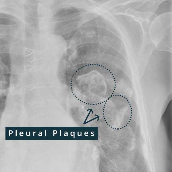
Asbestos related pleural and parenchymal fibrosis. Plain chest radiography is usually the first line radiologic examination and may demonstrate circumferential pleural thickening pleural effusions and lung nodules or. Mesothelioma also known as malignant mesothelioma is an aggressive malignant tumour of the mesothelium.

A comparison of ct and chest radiography. It shares epidemiological and pathological features with but is less common than its pleural counterpart which is described in detail in the general article on mesothelioma. Most tumors arise from the pleura and so this article will focus on pleural mesothelioma.

Involving any of the ipsilateral pleural surfaces with at least one of the. The role of computed tomography scanning in the initial assessment and the follow up of malignant pleural mesothelioma. Mediastinal and diaphragmatic pleura visceral pleura.

Mesothelioma also known as malignant mesothelioma is an aggressive malignant tumor of the mesothelium. Detection with high resolution ct.


Randulfus40 Asbestosis Vs Mesothelioma Radiology


Mesothelioma Imaging And Diagnostics What To Look For Imaging Technology News


Pleural Plaques Causes Symptoms Diagnosis Treatment


Radiological Review Of Pleural Tumors Sureka B Thukral Bb Mittal Mk Mittal A Sinha M Indian J Radiol Imaging


Overview Of Treatment Related Complications In Malignant Pleural Mesothelioma Murphy Annals Of Translational Medicine


Mesothelioma Lung Cancer X Ray Stock Image C017 7776 Science Photo Library


Localized Malignant Pleural Sarcomatoid Mesothelioma Misdiagnosed As Benign Localized Fibrous Tumor Kim Journal Of Thoracic Disease

Ct Scan Mesothelioma Radiology

Ct scan if doctors suspect mesothelioma in their patients they may recommend various diagnostic tools including medical imaging procedures. Mesothelioma metastasis radiology ct scans are usually used for pleural mesothelioma and peritoneal mesothelioma but not for the pericardial type of the disease.


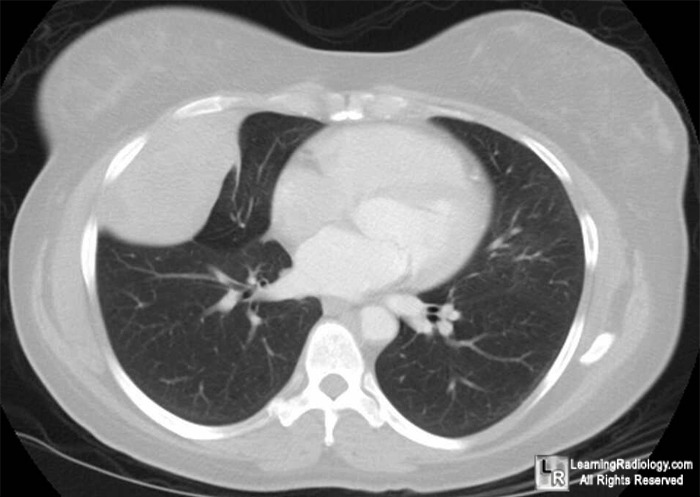
Learningradiology Localized Fibrous Tumor Pleura Solitary Benign Mesothelioma Pleural Fibroma Radiology

Staging of malignant pleural mesothelioma.

Ct scan mesothelioma radiology. It is generally considered too risky to use ct level radiation over the heart. Crossref medline google scholar. Comparison of ct and mr imaging.

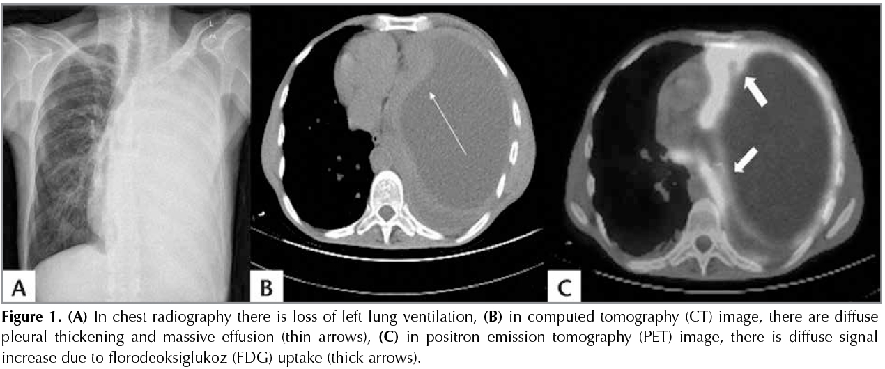
J digit imaging 2000. This image shows the extensive pleural thickening that is characteristic of mesothelioma effusion and reduction in the volume of the affected hemithorax. Malignant peritoneal mesothelioma is an uncommon primary tumor of the peritoneal lining.

It shares epidemiological and pathological features with but is less common than its pleural counterpart which is described in detail in the general article on mesothelioma. They often allow doctors to detect mesothelioma tumors including location sizing and potential spreading to aid in diagnosis and staging. Ct scans enable doctors to identify the stage of a tumor by exposing whether the tumor has spread to nearby tissues lymph nodes or to distant organs.

This imaging technology is less valuable in the diagnosis and staging of peritoneal mesothelioma but can be helpful in specific instances. The computed tomography scan is an important tool in this process because it can provide highly detailed information about the disease type location and metastasis. Other abdominal subtypes also discussed separately include.

Imaging scans are often a first step in a mesothelioma diagnosis. Resolution of pet scan images are relatively low hence the use of dual imaging combinations of pet and ct scans at most modern cancer centers. Initial chest radiography and chest computed tomography ct demonstrated circumferential lobulated pleural thickening involving the left lung with associated left lung volume loss figs 1 2whole body integrated positron emission tomography petct demonstrated circumferential pleural thickening surrounding the entire left lung with significant hypermetabolic.

21 heelan rt rusch vw begg cb panicek dm caravelli jf eisen c. X rays mri scans ct scans pet scans and ultrasounds are performed to detect abnormalities and potential causes of symptoms. Ct is most commonly used for imaging assessment of mesothelioma and sufficient for accurate staging of disease in most patients.

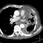
Pleural mass or nodular thickening of soft tissue attenuation tends to cause inward contraction of the hemithorax eg. Use of three dimensional spiral computed tomography imaging for staging and surgical planning of head and neck cancer. Computed tomography scan of a 58 year old patient with mesothelioma and shortness of breath.

Many doctors concur that ct scans provide the best imaging technology for scans of the abdomen and chest the areas most prone to the formation of mesothelioma.


Malignant Peritoneal Mesothelioma Radiology Reference Article Radiopaedia Org


Pleural Mesothelioma Dr Tinku Joseph


The Role Of Imaging In Malignant Pleural Mesothelioma An Update After The 2018 Bts Guidelines Clinical Radiology


Malignant Pleural Mesothelioma Evaluation With Ct Mr Imaging And Pet Radiographics


Malignant Peritoneal Mesothelioma Radiology Reference Article Radiopaedia Org


Https Encrypted Tbn0 Gstatic Com Images Q Tbn 3aand9gcrfu2kopu H3lo8g6sgjdfghewrsl9zt9czknshwv2eyugmcgbv Usqp Cau


Primary Peritoneal Mesothelioma Radiology Case Radiopaedia Org