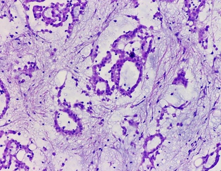
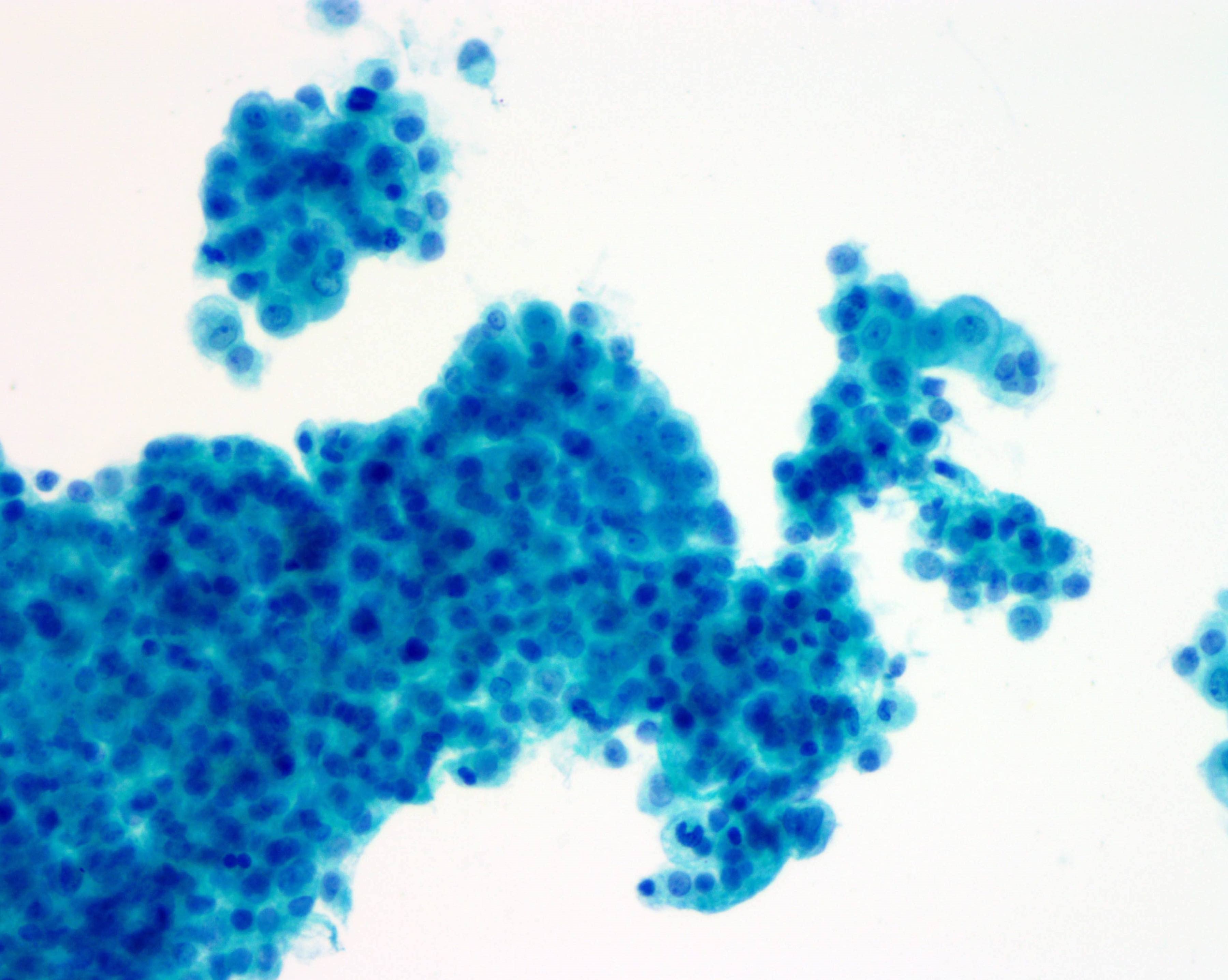
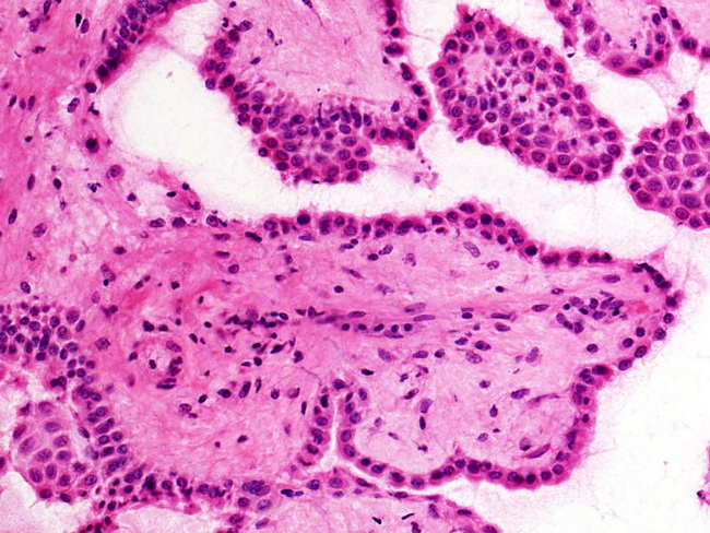
Tag Archives: malignant mesothelioma cytology

Malignant Mesothelioma Cytology

In this distinction pax8 expression has been regarded as a specific marker of serous carcinoma. For mesothelioma patients this fluid most commonly is either pleural effusion in pleural malignant mesothelioma patients or peritoneal effusion for those with mesothelioma of the peritoneum.
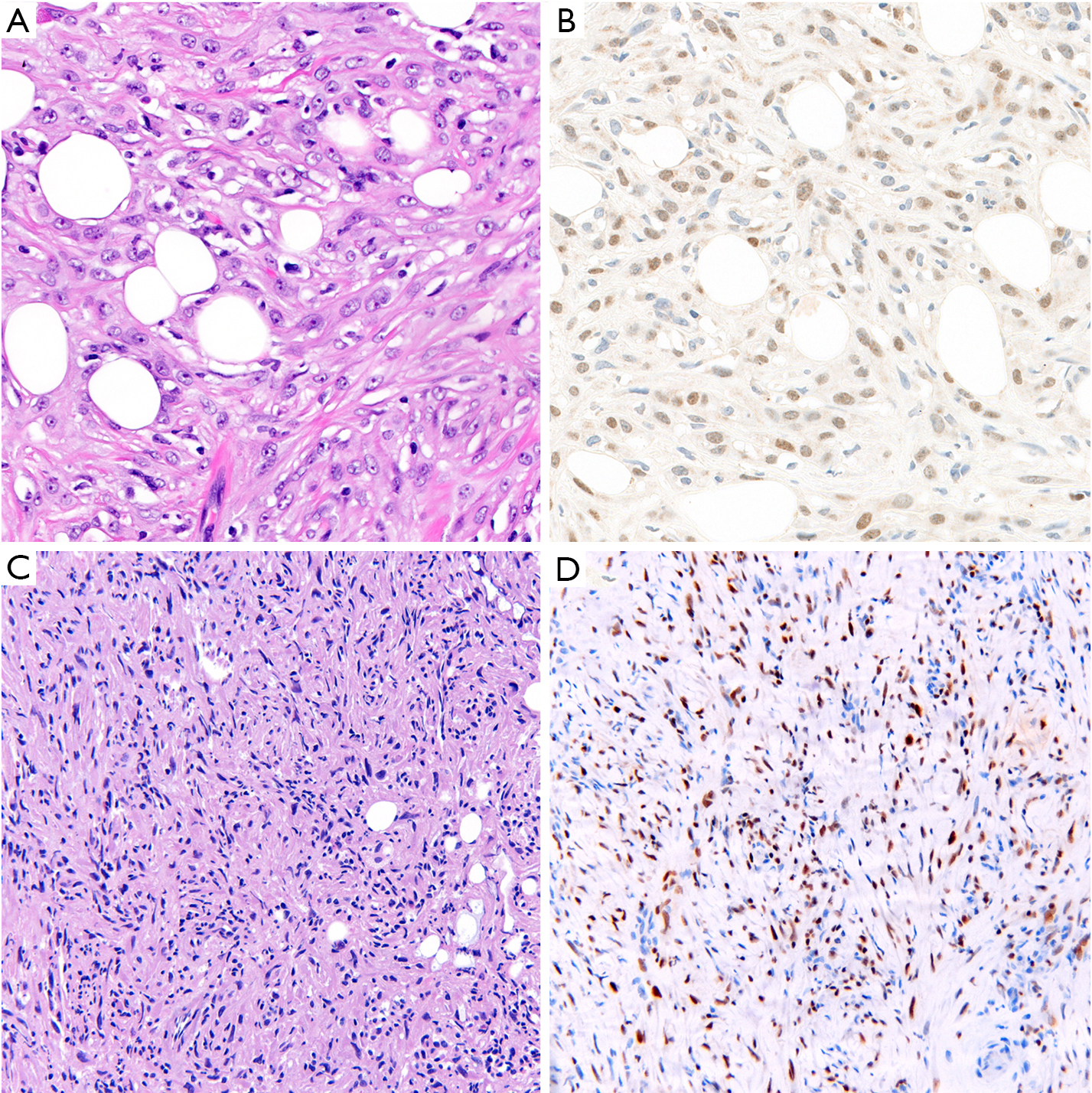


Application Of Immunohistochemistry In Diagnosis And Management Of Malignant Mesothelioma Chapel Translational Lung Cancer Research

Signs and symptoms of mesothelioma may.

Malignant mesothelioma cytology. The most common area affected is the lining of the lungs and chest wall. Mesothelioma specialists tend to have the best results when drawing cytology samples as they know exactly which parts of the body are most likely to collect mesothelioma cells. The tumour grows as multiple plaques and large nodules on the serosal surface.

Mesothelioma cytology or mesothelioma cytopathology is the study of cells for the presence of mesothelioma. Most patients have an effusion at the time of presentation. Siddiqui fernando schmitt andrew churg proceedings of the american society of cytopathology companion session at the 2019 united states and canadian academy of pathology annual meeting part 2.

In an effort to estimate the practice at other institutions a survey was disseminated regarding cytologic diagnosis of mm. Less commonly the lining of the abdomen and rarely the sac surrounding the heart or the sac surrounding the testis may be affected. Malignant mesothelioma arises most commonly in the pleura and rarely in the peritoneum.

It is a part of mesothelioma pathology which is the study of tissue or fluid to determine if this cancer exists. Malignant mesothelioma in cytology how far should we go. At the study institution northwestern university a primary diagnosis of mm is made on fluid cytology specimens.

The occurrence of this tumour is related to asbestos exposure. Follow the guidelines common sense clinicalimaging findings and remember behind every glass slide there is a human being and soon. Mesothelioma is a type of cancer that develops from the thin layer of tissue that covers many of the internal organs known as the mesothelium.

Mesothelioma cytology examines single cells often through analysis of bodily fluids. Distinguishing malignant peritoneal mesothelioma mpm from serous carcinoma involving the peritoneum remains a diagnostic challenge particularly in small biopsy and cytology specimens. Effusion cytology with focus on theranostics and diagnosis of malignant mesothelioma journal of the american society of cytopathology 10.

Never make a dx on cytology dx of mesothelioma on cytology piece of cake. Patients who suspect a mesothelioma diagnosis should therefore seek out the help and expertise of a qualified mesothelioma specialist. The diagnosis of malignant mesothelioma mm in effusion specimens is controversial.

In addition bap.
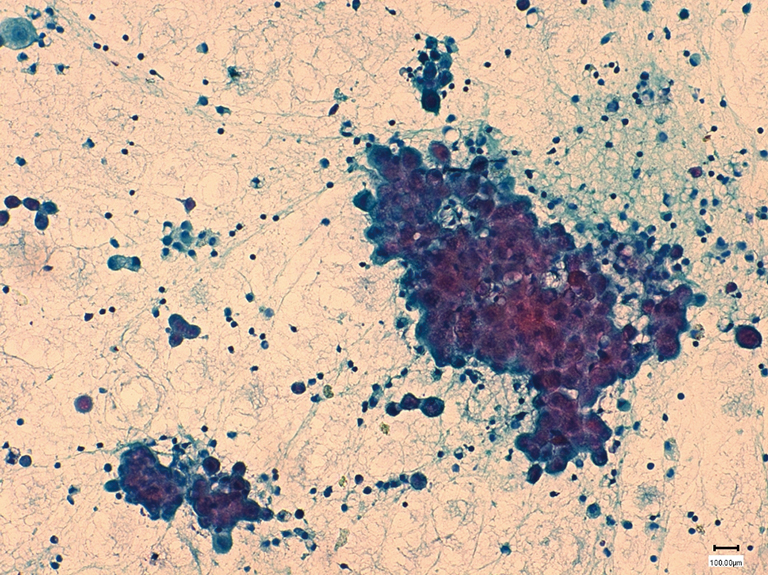


The Pathological And Molecular Diagnosis Of Malignant Pleural Mesothelioma A Literature Review Ali Journal Of Thoracic Disease


Malignant Mesothelioma Cytology


Cytomorphological Profile Of Neoplastic Effusions An Audit Of 10 Years With Emphasis On Uncommonly Encountered Malignancies Gupta S Sodhani P Jain S J Can Res Ther
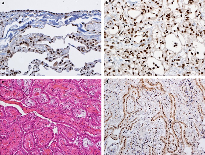


Bap1 Brca1 Associated Protein 1 Is A Highly Specific Marker For Differentiating Mesothelioma From Reactive Mesothelial Proliferations Modern Pathology
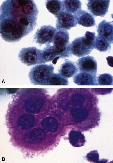


Malignant And Borderline Mesothelial Tumors Of The Pleura Thoracic Key


Https Encrypted Tbn0 Gstatic Com Images Q Tbn 3aand9gcsfu Mxmneu Gwnfgj0lkypoqodnbul8473zuq95ckzmrca Tnd Usqp Cau
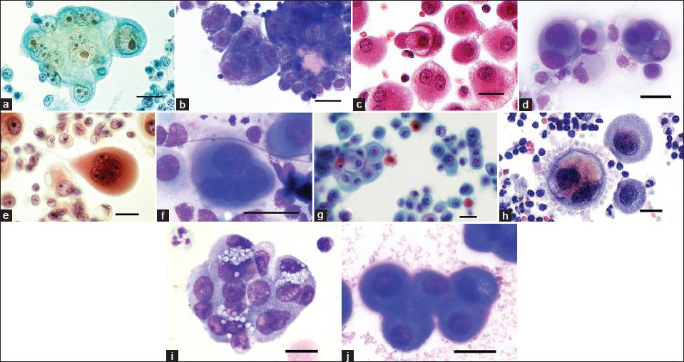


Guidelines For Cytopathologic Diagnosis Of Epithelioid And Mixed Type Malignant Mesothelioma Complementary Statement From The International Mesothelioma Interest Group Also Endorsed By The International Academy Of Cytology And The Papanicolaou Society Of
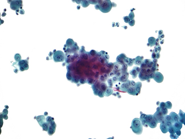
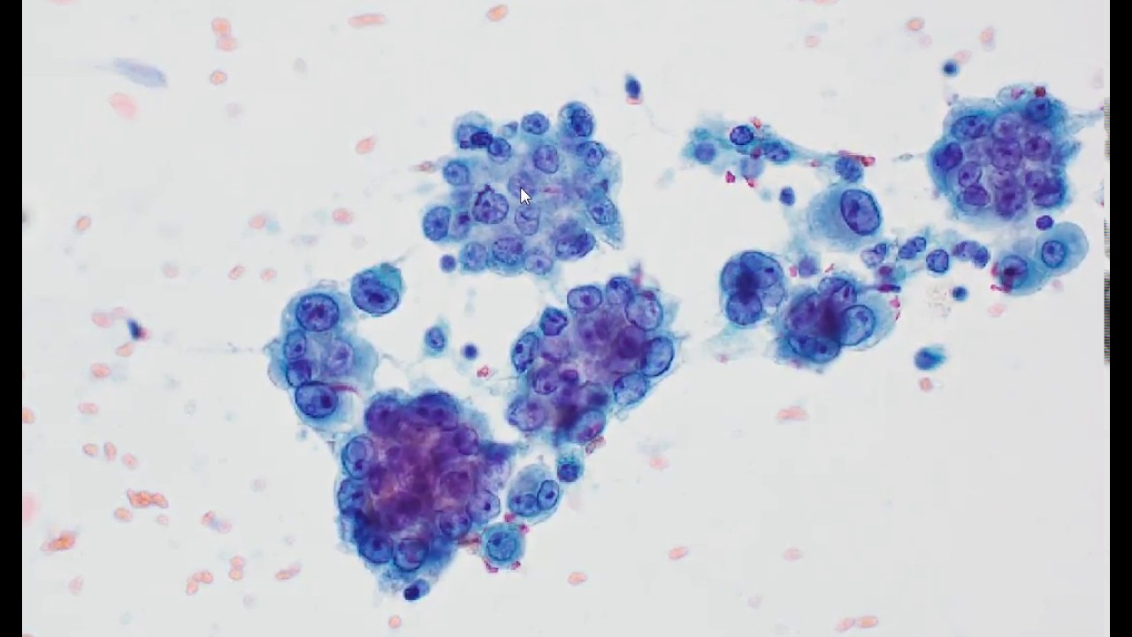
Mesothelioma Cytology

It deals with pericardial fluid peritoneal fluid and pleural fluid. Signs and symptoms of mesothelioma may.


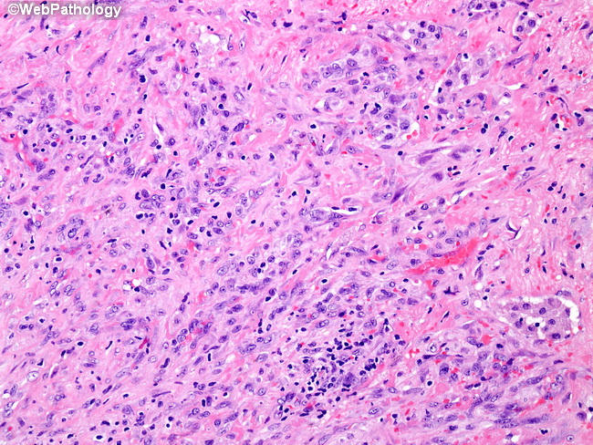
Webpathology Com A Collection Of Surgical Pathology Images

This fact is due to tissue samples being more reliable than fluid samples.

Mesothelioma cytology. It is a part of mesothelioma pathology which is the study of tissue or fluid to determine if this cancer exists. One study published on uptodate compared the accuracy of tissue biopsies and fluid samples. Mesothelioma cytology unlike imaging tests cytology is a pathological means to diagnosing malignant mesothelioma in patients consisting of a fluid biopsy of abnormalities previously discovered.

Mesothelioma cytology or cytopathology goes hand in hand with histology. However there is still a small risk of tumor cell seeding microscopic spreading of cancer due to cells moving during a biopsy during cytologic procedures. A look at how the cancer will advance.

Mesothelioma histology is more trusted than cytology for pathologically diagnosing this disease. By forgoing a tissue biopsy there is less risk of morbidity of the patient. Less commonly the lining of the abdomen and rarely the sac surrounding the heart or the sac surrounding the testis may be affected.

The article deals with cytopathology specimens from spaces lined with mesothelium ie. The most common area affected is the lining of the lungs and chest wall. While the histological determination will focus more on the cell type the cytology research will dive deeper into how these cells are forming interacting and spreading in the body.

Mesothelial cytopathology is a large part of cytopathology. Mesothelioma cytology or mesothelioma cytopathology is the study of cells for the presence of mesothelioma. Mesothelioma is a type of cancer that develops from the thin layer of tissue that covers many of the internal organs known as the mesothelium.

Cytology is often one of the first tests used in the diagnosis of mesothelioma after imaging tests reveal the possibility of cancer. Mesothelioma cytology is a useful tool that enables analysis without necessitating a tissue biopsy. Carlos wm bedrossian an update on pleuropulmonary cytopathology.

Once cytology is performed the cells from the sample are examined to arrive at a diagnosis. Cytology or fluid biopsy is a pathology specialty which studies disease by looking at the appearance and behavior of cells in bodily fluids. Mesothelioma cytology mesothelioma cytology.

Cytological diagnosis of mesothelioma and molecular cytology of lung cancer with an historical perspective diagnostic cytopathology 101002dc23298 43 7 513 526 2015.


Figure 2 A B Neoplastic Cells Arrows Intermixed With Reactive Mesothelial Cells Note The Eccentric Location Of The Nuclei Of Neoplastic Cells Kletki Krovi


Moving Toward An Earlier Diagnosis Of Mesothelioma


Cytological Profile Of A Case Of Malignant Mesothelioma Of Pleura With Supraclavicular Lymph Node Metastasis Abstract Europe Pmc


Https Onlinelibrary Wiley Com Doi Pdf 10 1002 Cncy 21342


Mesothelioma Dog


Https Encrypted Tbn0 Gstatic Com Images Q Tbn 3aand9gcsbgvpsjkfmxbyb2dtepoaktmrusji5uunvmadtoap8gksrx8cp Usqp Cau


The Value Of Brca 1 Associated Protein 1 Expression And Cyclin Dependent Kinase Inhibitor 2a Deletion To Distinguish Peritoneal Malignant Mesothelioma From Peritoneal Location Of Carcinoma In Effusion Cytology Specimens Althakfi 2020

Malignant Mesothelioma


Malignant Mesothelioma Springerlink


Histopathology Images Of Malignant Mesothelioma By Pathpedia Com


Malignant Mesothelioma Mm American Urological Association


Histopathology Images Of Malignant Mesothelioma By Pathpedia Com


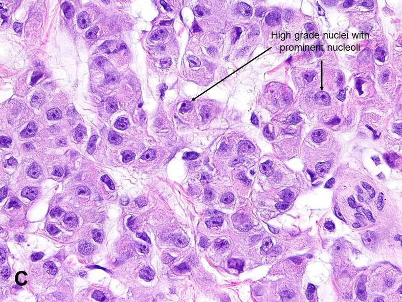
A Epithelioid Malignant Mesothelioma Grading Nuclear Grade I With


Webpathology Com A Collection Of Surgical Pathology Images


A Subset Of Malignant Mesothelioma Tumors Retain Osteogenic


Texas Asbestos Lawyer 12 Questions Answered About Malignant

Malignant Mesothelioma Cytology

Mesothelioma cytology is referred to as cytopathology because it is included in the field of pathologythe study of the behavior of diseases. Peritoneal mesothelioma is the second most common form of the disease accounting for less than 30 of all cases.


Fine Needle Aspiration Biopsy Of Metastatic Malignant Mesothelioma

Malignant mesothelioma arises most commonly in the pleura and rarely in the peritoneum.

Malignant mesothelioma cytology. Most patients have an effusion at the time of presentation. Signs and symptoms of mesothelioma may. Similar to pleural mesothelioma the disease also causes a build up of excess fluid in the abdominal cavity.

To assess the role of effusion cytology in mesothelioma diagnosis we conducted an audit of pleural fluid cytology results over a 20 year period 1988. Less commonly the lining of the abdomen and rarely the sac surrounding the heart or the sac surrounding the testis may be affected. The tumour grows as multiple plaques and large nodules on the serosal surface.

Mesothelioma cytology evaluates the behavior of mesothelioma cells. Other common causes include pleural mesothelioma and lymphoma. The cytology of malignant mesothelioma.

Mesothelioma cytology thus is a procedure to rate the behavior of mesothelioma cells which are in an abnormal fluid build up. The incidence has increased in the past two decades but still it is a rare tumor. That is our findings clearly indicate that negative staining for bap1 in effusion cytology supports a diagnosis of mesothelioma.

Malignant cells form in the peritoneum affecting the abdomen bowel liver and spleen. The most common area affected is the lining of the lungs and chest wall. Etiology of all forms of mesothelioma is strongly associated with.

Mesothelioma is a type of cancer that develops from the thin layer of tissue that covers many of the internal organs known as the mesothelium. However benign pleural effusions greatly outnumber effusions due to mesothelioma and therefore for an ancillary marker of malignant mesothelioma to have real clinical utility it must be highly specific. Malignant mesothelioma mm is a rare but rapidly fatal and aggressive tumor of the pleura and peritoneum with limited knowledge of its natural history.

If you think you or someone you adore might be in danger for mesothelioma you might wish to consider cytology as a choice for diagnosis or screening. Lung cancer and breast cancer account for about 50 65 of malignant pleural effusions. This process is useful for doctors in diagnosing researching and understanding mesothelioma.

Malignant pleural effusion is a condition in which cancer causes an abnormal amount of fluid to collect between the thin layers of tissue lining the outside of the lung and the wall of the chest cavity. The occurrence of this tumour is related to asbestos exposure.


Clinical Data And P16 Fish Results In 20 Cases Of Malignant Pleural


A Case Of Malignant Mesothelioma Showing Increased Cellularity


Pathology Walker Auto Tweet Bot On Twitter Malignant Mesothelioma


Pathology Outlines Epithelioid Mesothelioma


Representative Photomicrographs Of Cases Of A And C Malignant


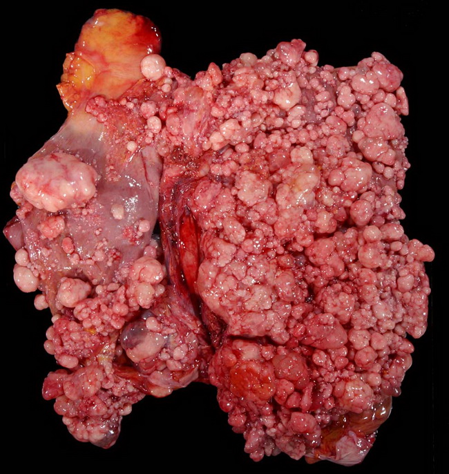
Omental Epithelial Malignant Mesothelioma Tumor Forms Two Small


Pathology Outlines Epithelioid Mesothelioma

Mesothelioma Cytology


Dinesh Rakheja On Twitter Impressed That Smlungpathguy Can


Guidelines For Cytopathologic Diagnosis Of Epithelioid And Mixed


Accme Disclosures


02 Presentations Ii Vs 14 4 Mb 3 30 08


Mesothelioma Cytology Youtube


Webpathology Com A Collection Of Surgical Pathology Images


Figure 1 From Cytological Diagnosis Of Malignant Mesothelioma


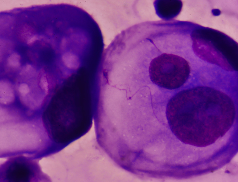
File Mesothelioma Cytology 3 Small Gif Wikimedia Commons