Tag Archives: malignant pleural mesothelioma ct

Malignant Pleural Mesothelioma Ct

Mediastinal and diaphragmatic pleura visceral pleura. Involving any of the ipsilateral pleural surfaces with at least one of the.


Mesothelioma Wikipedia

Malignant pleural mesothelioma is a rare tumor.
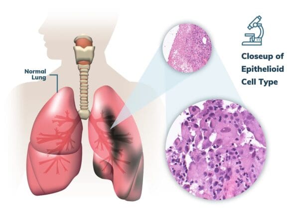
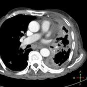
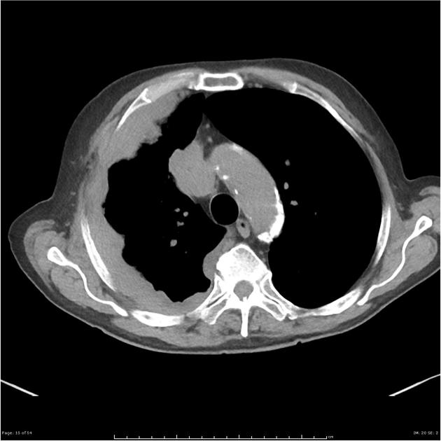
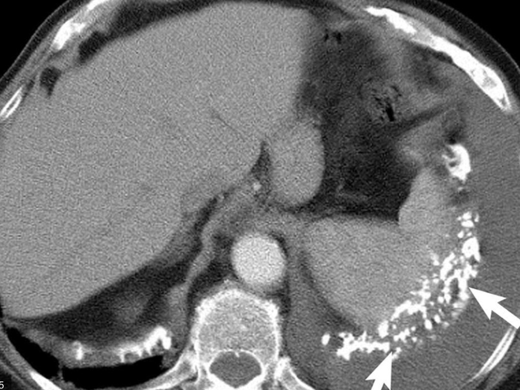
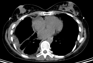
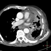
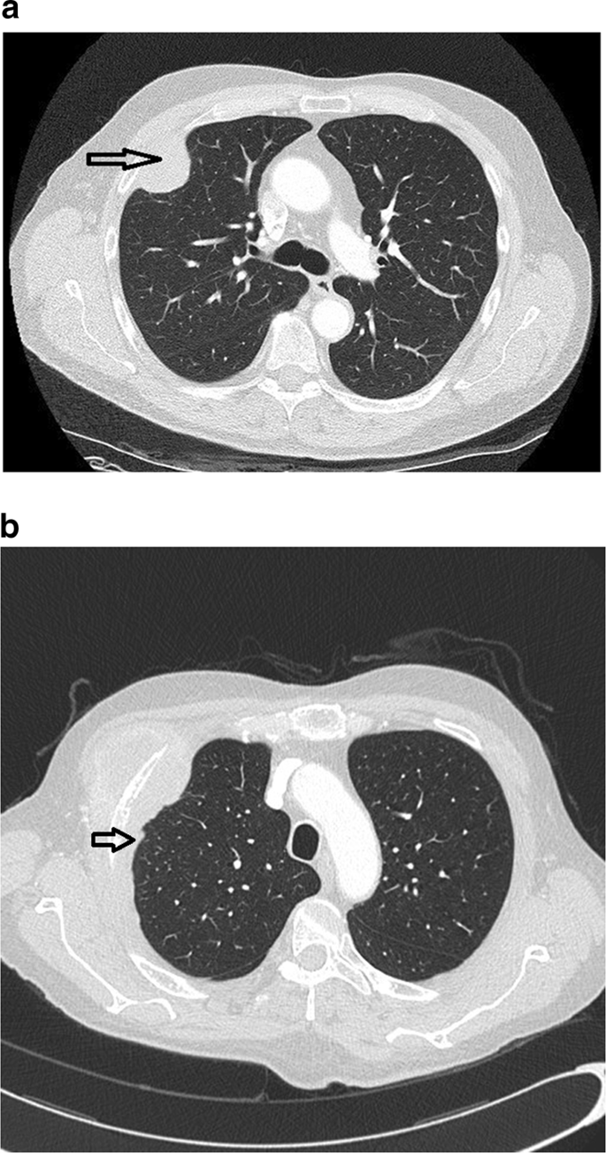
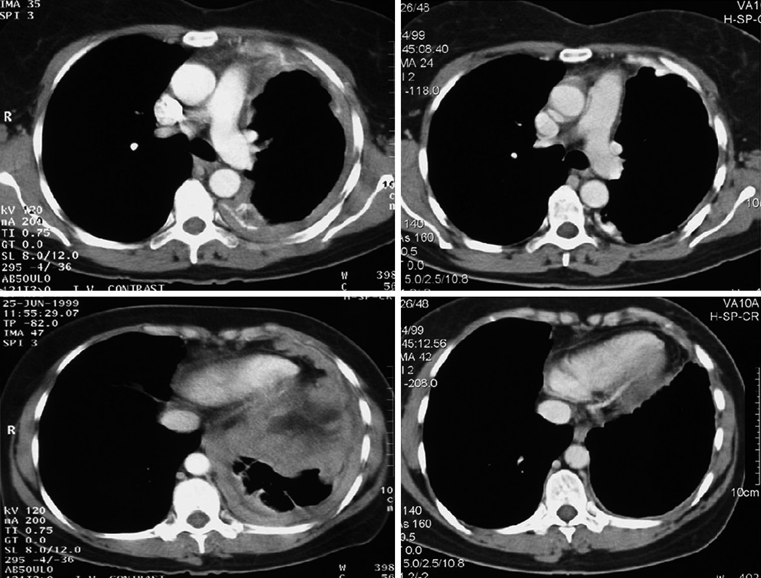
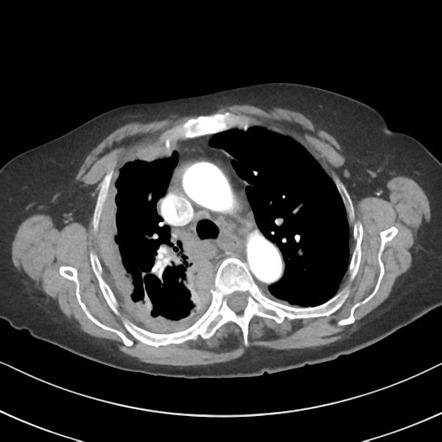
Malignant pleural mesothelioma ct. The tumor originates from cells of the visceral or parietal pleural and is linked to asbestos exposure with a median latency of 446 years due to the latency between exposure and onset of mesothelioma and the ongoing use of asbestos in parts of the world the incidence is expected to rise. Although the chest film findings of pleural mesothelioma are well described there are few descriptions of the findings of computed tomography ct. Computed tomography is the primary imaging modality used for the diagnosis and staging of mpm.

This condition is usually associated with occupational exposure to asbestoswagner et al connected asbestos to mesothelioma in a classic 1960 study of 33 patients with mesothelioma who were exposed to asbestos in a mining area in south africas north western cape province. Alexander e clark ra colley dp mitchell se. Ct of malignant pleural mesothelioma.

Below is the eighth edition of the tnm staging system for malignant pleural mesothelioma which was published in 2018 1. Malignant pleural mesothelioma mpm is a rare and aggressive form of cancer that originates in the pleura within the chest cavity. Involving ipsilateral parietal pleura inc.

Additionally well designed clinical trials are needed to determine the effectiveness of ct screening in populations exposed to asbestos. This report describes the ct findings in five cases of pleural. Discuss the advan tages and limitations of ct mr imaging and pet in the diag nosis and staging of mpm.

Ct manifestations in 50 cases. Magnetic resonance mr imaging and more recently positron emission tomography pet have emerged as modalities. Imaging plays an essential role in the evaluation of malignant pleural mesothelioma mpm.

The task force recommends large international epidemiological studies to determine the relationship between pleural plaques and malignant pleural mesothelioma. Most tumors arise from the pleura and so this article will focus on pleural mesothelioma. Describe key fea tures of mpm at ct mr imaging and pet.

Evaluation with ct mr imaging and pet1 learning objectives for test 4 after reading this article and taking the test the reader will be able to. 15 kawashima a libshitz hi. Crossref medline google scholar.

Given the presence of the mesothelium in different parts of the body mesothelioma can arise in various locations 17. Primary tumor cannot be assessed. No evidence of primary tumor.

Ajr am j roentgenol 1990. Pleural mesothelioma 90 covered in this article. Mesothelioma is a malignant neoplasm originating from pleural or peritoneal surfaces.

Mesothelioma also known as malignant mesothelioma is an aggressive malignant tumor of the mesothelium. The pleura is a thin cellular lining than envelops both the lung termed the visceral pleura and chest wall the parietal pleura as well as the diaphragm and the heart and major blood vessels in the central portion of the chest. Malignant pleural mesothelioma mpm is an aggressive thoracic malignancy with a dismal prognosis.


Mesothelioma Radiology Case Radiopaedia Org


Computed Tomography Ct Showing Advancedstage Pleural Mesothelioma Download Scientific Diagram


Https Pubs Rsna Org Doi Pdf 10 1148 Rg 241035058


Iranian Journal Of Radiology Malignant Mesothelioma Versus Metastatic Carcinoma Of The Pleura A Ct Challenge


Quick Take Prophylactic Irradiation Of Tracts In Patients With Malignant Pleural Mesothelioma An Open Label Multicenter Phase Iii Randomized Trial 2 Minute Medicine


Pleural Mesothelioma Stages Treatment Prognosis


Radiological Review Of Pleural Tumors Sureka B Thukral Bb Mittal Mk Mittal A Sinha M Indian J Radiol Imaging
Malignant Mesothelioma Ct

It shares epidemiological and pathological features with but is less common than its pleural counterpart which is described in detail in the general article on mesothelioma. Given the presence of the mesothelium in different parts of the body mesothelioma can arise in various locations 17.


Localized Malignant Pleural Sarcomatoid Mesothelioma Misdiagnosed As Benign Localized Fibrous Tumor Kim Journal Of Thoracic Disease

Malignant peritoneal mesothelioma is an uncommon primary tumor of the peritoneal lining.

Malignant mesothelioma ct. Involving ipsilateral parietal pleura inc. Pleural mesothelioma 90 covered in this article. Mesothelioma also known as malignant mesothelioma is an aggressive malignant tumour of the mesothelium.

Computed tomography ct is considered the standard for first line imaging of mpem diagnosis. Malignant peritoneal mesothelioma mpem is a rare cancer of the mesothelial cells of the peritoneum. No evidence of primary tumor.

Plain radiographic and ct findings alone are nonspecific but in the presence of a long history of asbestos exposure they can be highly suggestive of malignant mesothelioma. Below is the eighth edition of the tnm staging system for malignant pleural mesothelioma which was published in 2018 1. Other abdominal subtypes also discussed separately include.

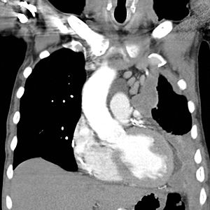
Ct scan of a patient with mesothelioma coronal section the section follows the plane that divides the body in a front and a back half. Pleural mesothelioma 90 covered in this article. Malignant deciduoid mesothelioma a rare phenotype of epithelioid mesothelioma arises more commonly from the peritoneum of young women but is also from the pleura of elderly people.

Primary tumor cannot be assessed. Ajr am j roentgenol 1990. Plain chest radiography is usually the first line radiologic examination and may demonstrate circumferential pleural thickening pleural effusions and lung nodules or.

This condition is usually associated with occupational exposure to asbestoswagner et al connected asbestos to mesothelioma in a classic 1960 study of 33 patients with mesothelioma who were exposed to asbestos in a mining area in south africas north western cape province. Mesothelioma also known as malignant mesothelioma is an aggressive malignant tumor of the mesothelium. Mediastinal and diaphragmatic pleura visceral pleura.

Given the presence of the mesothelium in different parts of the body mesothelioma can arise in various locations 17. In the current report the authors describe an unusual case of peritoneal malignant epithelioid mesothelioma with rare deciduoid phenotype demonstrated with 67 ga. Involving any of the ipsilateral pleural surfaces with at least one of the.

Most tumors arise from the pleura and so this article will focus on pleural mesothelioma. Crossref medline google scholar. Most tumours arise from the pleura and so this article will focus on pleural mesothelioma.

The mesothelioma is indicated by yellow arrows the central pleural effusion fluid collection is marked with a yellow star. 1 right lung 2 spine 3 left lung 4 ribs 5 descending part of the aorta 6 spleen 7 left kidney 8. Mesothelioma is a malignant neoplasm originating from pleural or peritoneal surfaces.

15 kawashima a libshitz hi. Ct manifestations in 50 cases.


Mesothelioma Radiology Reference Article Radiopaedia Org


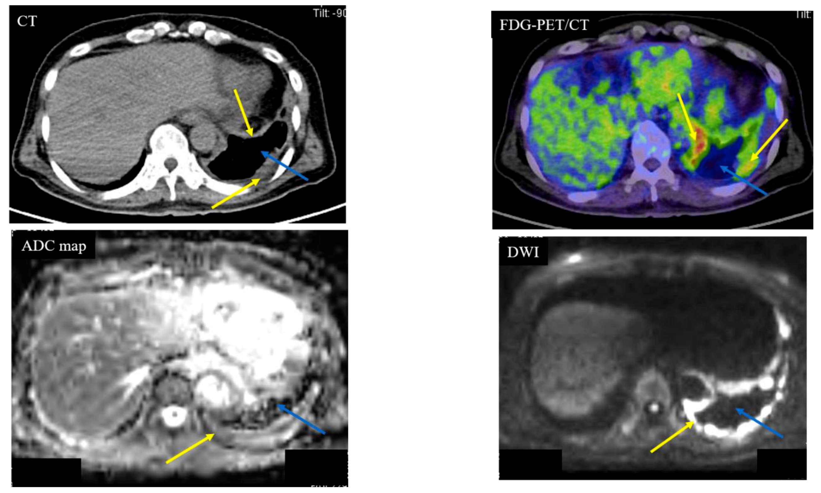
Cancers Free Full Text Diffusion Weighted Imaging Can Differentiate Between Malignant And Benign Pleural Diseases Html


Ct Scanning Not Reliable For Correct Diagnosis Of Pleural Mesothelioma


Figure 4 Malignant Mesothelioma Presenting As A Gradually Enlarging Pneumothorax


Heritable Mutations May Indicate Responsiveness To Platinum Chemotherapy In Malignant Mesothelioma Center For Cancer Research National Cancer Institute


Multimodality Imaging For Characterization Classification And Staging Of Malignant Pleural Mesothelioma Radiographics


Mesothelioma Of The Tunica Vaginalis In A Patient With Giant Hydrocele

Mesothelioma Ct

The connecticut mesothelioma victims centers services are available to people with mesothelioma or asbestos exposure lung cancer in new london groton bridgeport new haven hartford stamford. Ct demonstrates similar nonspecific findings.


Https Encrypted Tbn0 Gstatic Com Images Q Tbn 3aand9gcqm2wnxdnjwxjiarbr9ietjmckxbf5eolhcrkbcawdyim9zjp0n Usqp Cau

Crossref medline google scholar.

Mesothelioma ct. Ct scan of a patient with mesothelioma coronal section the section follows the plane that divides the body in a front and a back half. Ct manifestations in 50 cases. Ajr am j roentgenol 1990.

1 right lung 2 spine 3 left lung 4 ribs 5 descending part of the aorta 6 spleen 7 left kidney 8. Additional evidence of asbestos exposure such as calcified or noncalcified pleural plaques may be evident. Mesothelioma also known as malignant mesothelioma is an aggressive malignant tumor of the mesothelium.

This scan also can show if it has spread to lymph nodes or other organs. Diagnostic ct scan for mesothelioma. However with a history of significant asbestos exposure a diagnosis of mesothelioma can be suggested.

Mesothelioma is a malignant neoplasm originating from pleural or peritoneal surfaces. Ct stands for computerized tomography. In addition to the previously mentioned findings ct can also be useful in demonstrating invasion of the tumor into the mediastinum chest wall diaphragm and pericardium which aids in disease.

Ct scans can help physicians to determine the stage of your tumor. When a person goes to the doctor with early signs of malignant pleural mesothelioma a ct scan of the chest is often the next step in making a diagnosis. 15 kawashima a libshitz hi.

Appearances of asbestosis vary with the duration and severity of the condition. A new technique called ct perfusion can show if cancer cells are spreading in the bloodstream. Given the presence of the mesothelium in different parts of the body mesothelioma can arise in various locations 17.

Ct scans create a 3 dimensional image of internal organs making it easier to spot mesothelioma tumors. Pleural mesothelioma 90 covered in this article. The mesothelioma is indicated by yellow arrows the central pleural effusion fluid collection is marked with a yellow star.

More is better a new report out of the uk suggests that a thorough diagnostic ct scan for pleural mesothelioma should include images of the abdomen and pelvis as well as the chest. This condition is usually associated with occupational exposure to asbestoswagner et al connected asbestos to mesothelioma in a classic 1960 study of 33 patients with mesothelioma who were exposed to asbestos in a mining area in south africas north western cape province. How a diagnostic ct scan works.

But a research team led by the university of bristol says a diagnostic ct scan that shows only the chest might miss mesothelioma tumors in other areas. Still many doctors say the ct scan is the best for the chest and abdomen which are where mesothelioma forms. Most tumors arise from the pleura and so this article will focus on pleural mesothelioma.

Chest radiograph may show irregular opacities with a fine reticular pattern. Ct scanning is an.


Epos C 0908


Mesothelioma What Is The Mesothelioma Mesothelioma Dugli


Mesothelioma Symptoms Diagnosis And Treatment Bmj Best Practice Us


Malignant Pleural Mesothelioma Evaluation With Ct Mr Imaging And Pet Radiographics


Https Pubs Rsna Org Doi Pdf 10 1148 Rg 241035058


Pdf Oesphageal Stenting For Palliation Of Malignant Mesothelioma


Chest Ct Presenting The Mesothelioma Download Scientific Diagram

Pleural Mesothelioma Ct

More is better a new report out of the uk suggests that a thorough diagnostic ct scan for pleural mesothelioma should include images of the abdomen and pelvis as well as the chest. Imaging plays an essential role in the evaluation of malignant pleural mesothelioma mpm.


Mesothelioma Symptoms Diagnosis And Treatment Bmj Best Practice

Mesothelioma also known as malignant mesothelioma is an aggressive malignant tumour of the mesothelium.

Pleural mesothelioma ct. Mesothelioma also known as malignant mesothelioma is an aggressive malignant tumor of the mesothelium. The tumor originates from cells of the visceral or parietal pleural and is linked to asbestos exposure with a median latency of 446 years due to the latency between exposure and onset of mesothelioma and the ongoing use of asbestos in parts of the world the incidence is expected to rise. Most tumors arise from the pleura and so this article will focus on pleural mesothelioma.

Ct manifestations in 50 cases. The mesothelioma is indicated by yellow arrows the central pleural effusion fluid collection is marked with a yellow star. Ct scan of a patient with mesothelioma coronal section the section follows the plane that divides the body in a front and a back half.

Ajr am j roentgenol 1990. Given the presence of the mesothelium in different parts of the body mesothelioma can arise in various locations 17. However with a history of significant asbestos exposure a diagnosis of mesothelioma can be suggested.

1 right lung 2 spine 3 left lung 4 ribs 5 descending part of the aorta 6 spleen 7 left kidney 8. Given the presence of the mesothelium in different parts of the body mesothelioma can arise in various locations 17. In addition to the previously mentioned findings ct can also be useful in demonstrating invasion of the tumor into the mediastinum chest wall diaphragm and pericardium which aids in disease.

Pleural mesothelioma is a cancer of the pleural cavity which is a thin membrane between the chest wall and lung cavity. The presence of pleural thickening is not enough to confirm a pleural mesothelioma diagnosis but it can be a sign of serious and significant asbestos exposure. When the disease is called malignant pleural mesothelioma this means it is cancerous and will spread.

15 kawashima a libshitz hi. Magnetic resonance mr imaging and more recently positron emission tomography pet have emerged as modalities. Malignant pleural mesothelioma mpm is an aggressive thoracic malignancy with a dismal prognosis.

Computed tomography is the primary imaging modality used for the diagnosis and staging of mpm. Diagnostic ct scan for mesothelioma. Because catching mesothelioma in an early stage can lead to a wider range of treatment options patients with asbestos related pleural thickening should seek regular cancer screening.

This specific disease makes up approximately 80 of all mesothelioma cases. Crossref medline google scholar. Most tumours arise from the pleura and so this article will focus on pleural mesothelioma.

Pleural mesothelioma 90 covered in this article. Pleural mesothelioma 90 covered in this article. Ct demonstrates similar nonspecific findings.

When a person goes to the doctor with early signs of malignant pleural mesothelioma a ct scan of the chest is often the next step in making a diagnosis.


Loihealth Malignant Pleural Mesothelioma Desktop Backgrounds For Free Hd Wallpaper Wallart Com


Localized Malignant Mesothelioma An Unusual And Poorly Characterized Neoplasm Of Serosal Origin Best Current Evidence From The Literature And The International Mesothelioma Panel Modern Pathology


Management Of Malignant Pleural Mesothelioma The European Experience Opitz Journal Of Thoracic Disease


Figure 2 From Pleural Mesothelioma In A Young Male Patient Semantic Scholar


Malignant Pleural Mesothelioma Evaluation With Ct Mr Imaging And Pet Radiographics


Mesothelioma What Is The Mesothelioma Mesothelioma Dugli


The Role Of 18f Fdg Pet Ct Integrated Imaging In Distinguishing Malignant From Benign Pleural Effusion