Tag Archives: mesothelial cells in pleural fluid

Mesothelial Cells In Pleural Fluid

Reactive pleural effusion showing mesothelial cells lymphocytes neutrophils and macrophages. The fluid can accumulate quickly if the mesothelial cells fail to function resulting in an unhealthy collection of fluids in the chest cavity.


Effusions Cytopathology Cellnetpathology

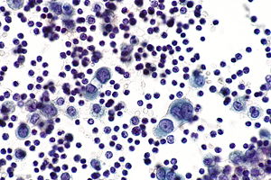
Numerous reactive mesothelial cells were present in only 12 of specimens examined.

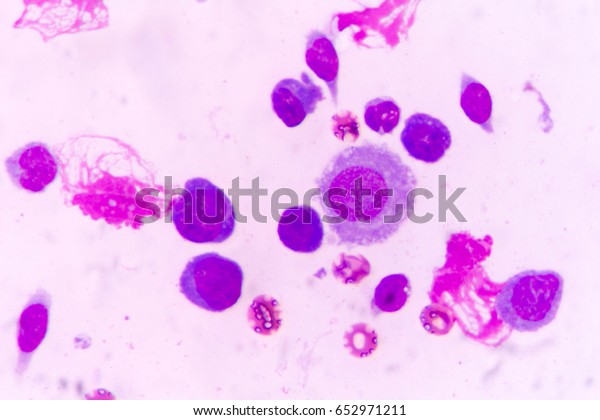
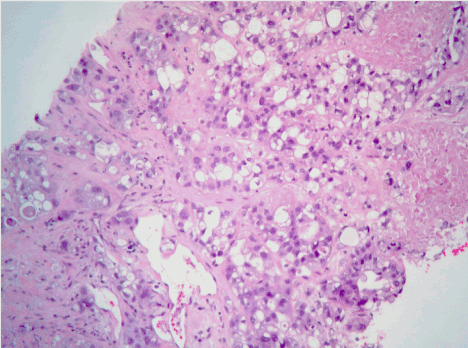
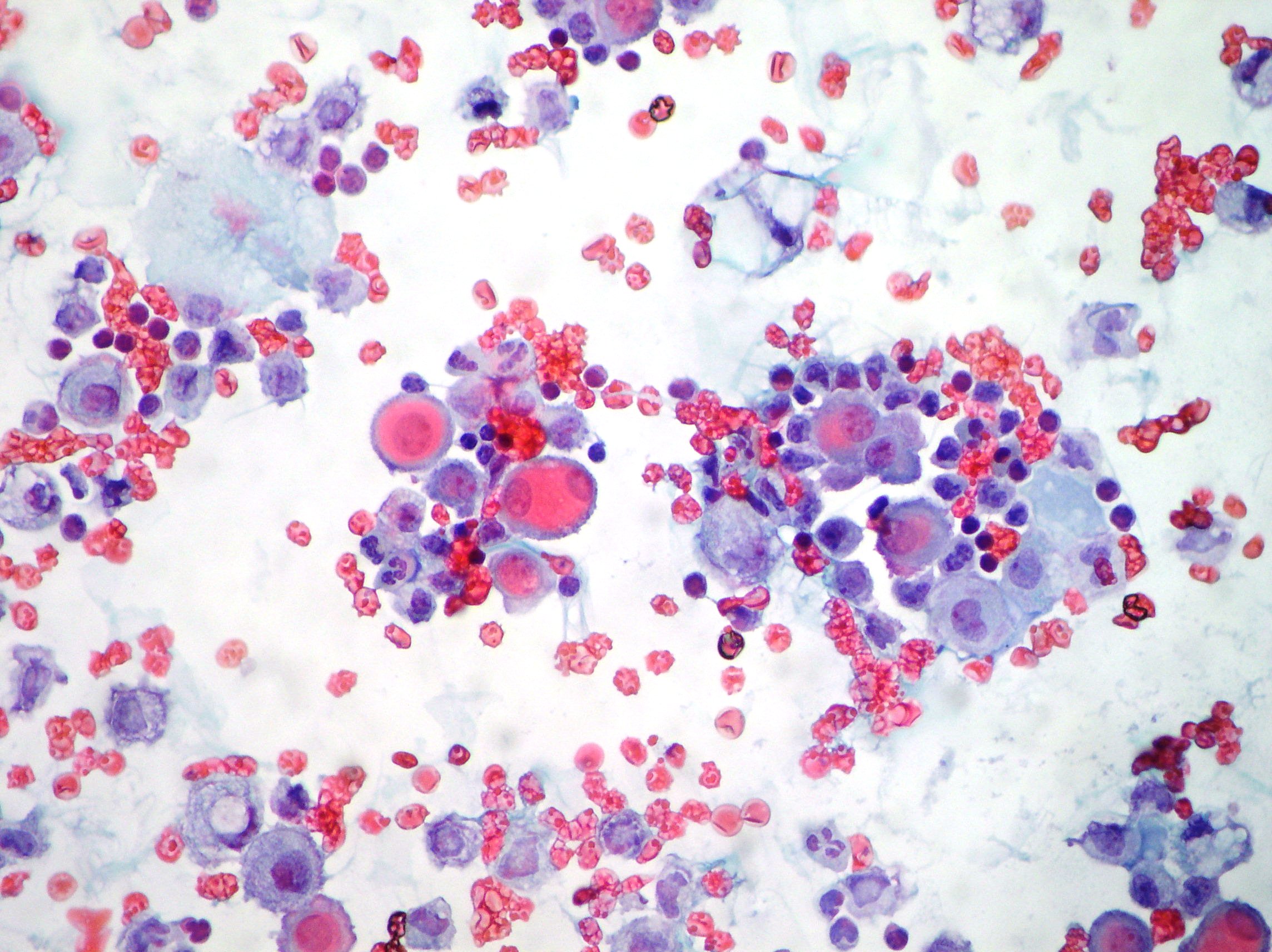
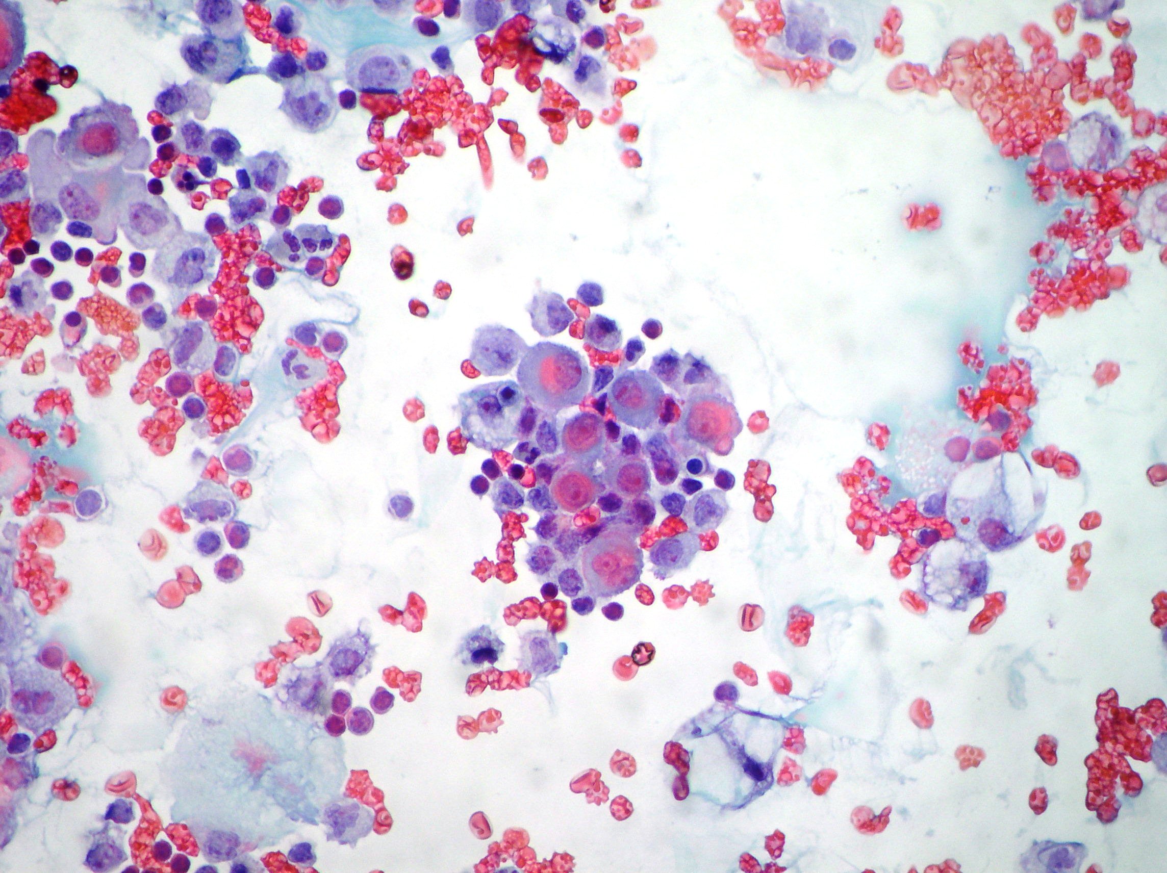
Mesothelial cells in pleural fluid. Reactive mesothelial cells present in a background of abundant lymphocytes. This has a large ddx. This condition can be due to the presence of a bacterial viral or fungal infection.

It can also be the result of trauma or the presence of metastatic tumor. Reactive mesothelial cells reactive mesothelial cells in pleural fluid reactive mesothelial cells are found when there is infection or inflammation present in a body cavity. Pleural effusion mesothelial cells pleural effusion.

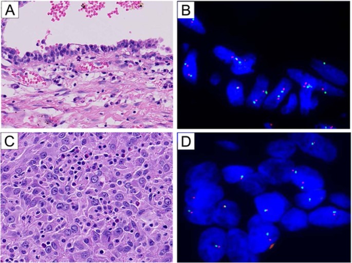
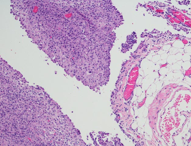
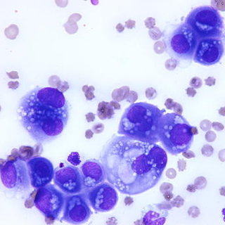
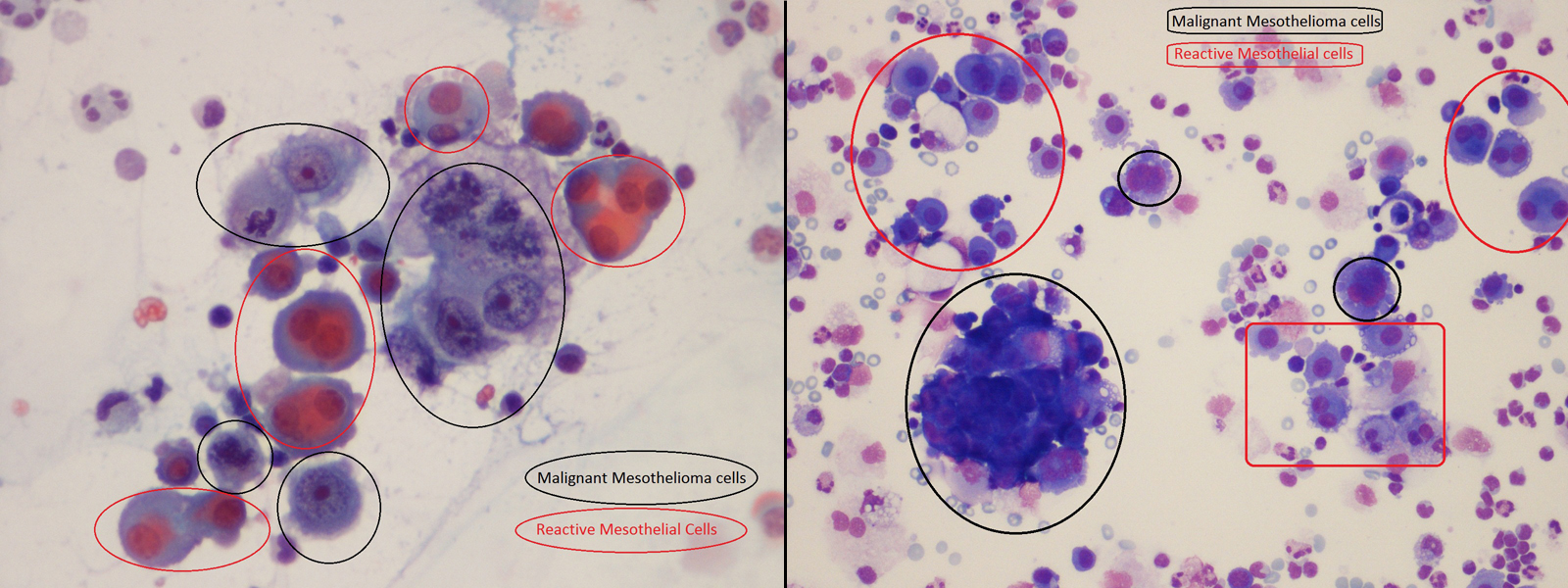
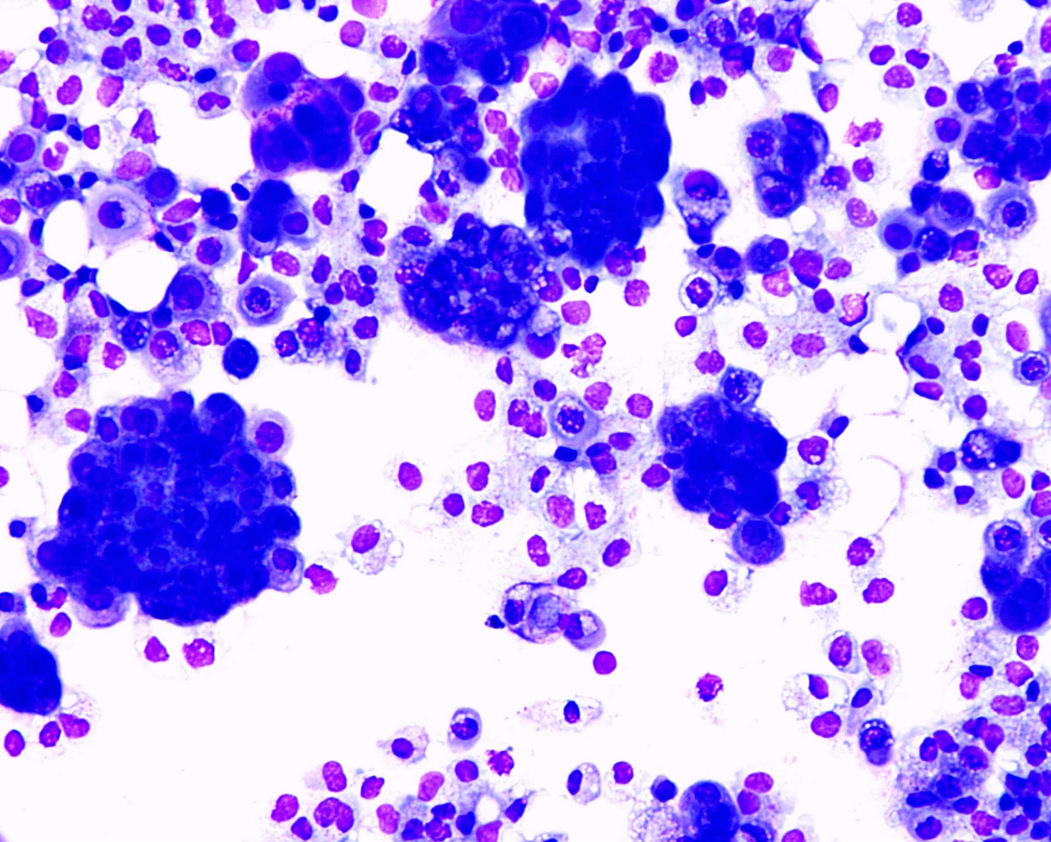
Negative for malignant cells. Pleural fluid right thoracentesis. Neoplastic transformation of mesothelial cells results in malignant mesothelioma an aggressive tumor especially the pleura.

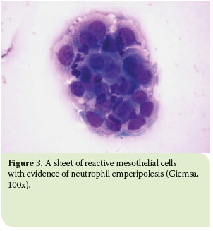
Immunocytochemistry test resulted positive for calretinin marker antibody papanicolaou x100 x200 mesothelial cells pleuric mesothelioma. Numerous mesothelial cells are seen in this pleural fluid from a dog with a transudative effusion with concurrent diapedesis of red blood cells or hemorrhage. Mesothelial cells are found in variable numbers in most effusions but their presence at greater than 5 of total nucleated cells makes a diagnosis of tb less likely.

This is known as pleural effusion. Reactive mesothelial cells can be found when there is an infection or an inflammatory response present in a body cavity. Actively dividing mesothelial cells can mimic an adenocarcinoma.

Pleural fluid for total white blood cell wbc count and differential cell count should be sent in an anticoagulated tube. A patient may develop shortness of breath and vague chest pains while the fluid experiences a buildup in the chest cavity these are two of the most common. Eighty five samples of pleural fluid obtained from 76 patients with biopsy proven tuberculous pleurisy were examined cytologically.

Specific diagnoses benign eosinophilic pleuritis general. Mesothelial cells in pleural fluid. Additional sampling should be considered within the clinical context.

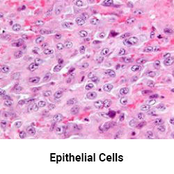
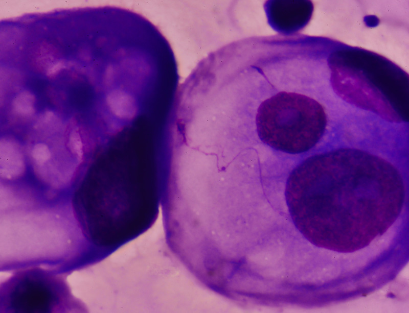
The mesothelial cells have central round nuclei with a moderate amount of light purple cytoplasm and a corona or fringe to the cytoplasmic borders. In contrast 653 of pleural fluid aspirates obtained from a control group of pati. There are certain cells that line the pleura the thin double layered lining which covers the lungs chest wall and diaphragm which are known as mesothelial cellsother than the pleura mesothelial cells also form a lining around the heart pericardium and the internal surface of the abdomen peritoneum.

Use of pleural fluid n. Trauma with air in the pleural cavity.


Http Www Cap Org Apps Docs Committees Hematology Educational Activities 2009 Cmb Pdf


Effusions Cytopathology Cellnetpathology


Pleural Fluid Smear Malignant Mesothelial Cells Lymphocytes Mgg Download Scientific Diagram


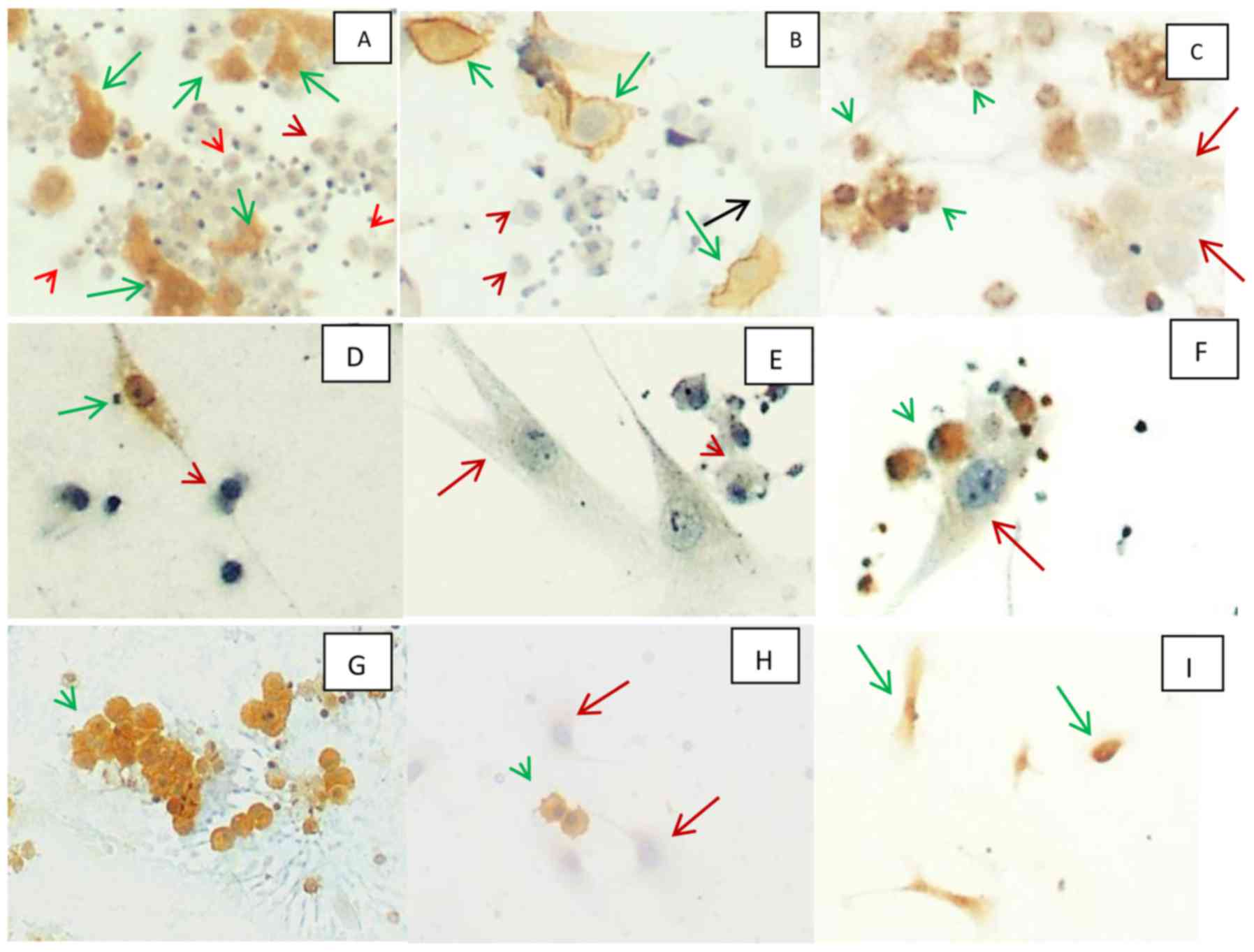
A Panel Of Markers For Identification Of Malignant And Non Malignant Cells In Culture From Effusions


View Image


Hjcam Iatrikh Zwwn Syntrofias Hellenic Journal Of Companion Animal Medicine Volume 6 Issue 1 2017 Pleural Effusion In The Cat A Focus On Laboratory Diagnosis


Http Www Cap Org Apps Docs Committees Hematology Educational Activities 2009 Cmb Pdf

Mesothelioma Cells

Mutations in mesothelial cells caused by asbestos exposure create these variations. Mesothelioma is a type of cancer that develops from the thin layer of tissue that covers many of the internal organs known as the mesothelium.


Hemizygous Loss Of Nf2 Detected By Fluorescence In Situ Hybridization Is Useful For The Diagnosis Of Malignant Pleural Mesothelioma Modern Pathology

Car t cell therapy is an emerging treatment and form of immunotherapy that uses a patients immune system to fight cancers like mesothelioma.

Mesothelioma cells. There are a number of factors that allow a doctor to diagnose the type of mesothelioma that you have and one of them is cell type. Mesothelial cells create a unique layer of paved like cells that line the bodys serious cavities and internal organs. The three major mesothelioma cell types are epithelioid sarcomatoid and biphasic.

Cancer starts when cells start to grow out of control. There are three main types of mesothelioma cells. Mesothelioma cell types explained.

Mesothelioma doctors consider cell type when creating a treatment plan because the cells respond differently to treatment. The most common area affected is the lining of the lungs and chest wall. In the case of mesothelioma when the mesothelial cells are exposed to asbestos fibers the cells ingest these fibers and prevent them from causing harm.

A mesothelioma patients treatment options and prognosis are directly affected by the different types of mesothelioma cells. There are different mesothelioma cells and each of them should be treated with a different treatment. Signs and symptoms of mesothelioma may.

Cells in nearly any part of the body can become cancer. For more information call us today at 888 204 2920. However if the asbestos exposure is continued over time such heavy volumes of asbestos can overwhelm the mesothelial cells.

Doctors will use cell type with mesothelioma type when determining viable treatment options and patient prognosis. The n body contains many different types of cells each of which has a specific function. About 60 percent of all mesothelioma a form of cancer caused by asbestos exposure cases are epithelioid.

The three mesothelioma cell types are epithelioid biphasic and sarcomatoid. The primary function of this layer termed the mesothelium is to provide a slippery non adhesive and protective surface. Malignant mesothelioma is cancer that starts in cells in the linings of certain parts of the body most commonly the linings of the chest or abdomen belly.

Epithelial sarcomatoid and biphasic. Car t cell therapy and mesothelioma. The epithelial mesothelioma cell type makes up more than 50 of all cases.

Researchers genetically modify t cells in a lab and inject them back into the patients body where they prevent or slow the spread of disease. Treatment and prognosis are affected by a patients. Less commonly the lining of the abdomen and rarely the sac surrounding the heart or the sac surrounding the testis may be affected.

Generally the types of mesothelioma cells can be significant in a patients prognosis and cancer treatment plan.


Multiple Sclerosis Drug Effectively Stops Mesothelioma Cells


Detection Of Malignant Mesothelioma Using Nuclear Structure Of Mesothelial Cells In Effusion Cytology Specimens Tosun 2015 Cytometry Part A Wiley Online Library


Ijms Free Full Text Experimental Model Of Human Malignant Mesothelioma In Athymic Mice


Benign Mesothelial Cells With Highly Irregular Nuclear Membranes And Download Scientific Diagram


Malignant Mesothelioma Cells Are Rapidly Sensitized To Trail Induced Apoptosis By Low Dose Anisomycin Via Bim Molecular Cancer Therapeutics


Mesothelioma Cell Types


Can You Identify Each Mesothelioma Cell Type Mesothelioma Guide

Mesothelioma Cells In Pleural Fluid

Cancer in the pleura may or may not cause symptoms. This is known as pleural effusion.


Case N 35 Pleural Effusion Mesothelioma Cytology Blog Site

Once in the pleura cancer cells may develop into one or multiple tumors or vice versa.

Mesothelioma cells in pleural fluid. In the current study researchers compared the traditional method of analyzing cancer cells in pleural fluid with a method that involves concentrating those scattered cells into a block that. This is due to medical intervention or tumor growth. Researchers from the moffitt cancer center have reported between 80 95 of pleural mesothelioma patients have pleural effusions.

While a pleural effusion may be a symptom of pleural mesothelioma itself the condition can also cause its own symptoms like breathlessness. A pleural effusion is an abnormal fluid buildup between the pleural layers. Looking for mesothelioma cells in pleural fluid can help doctors identify this asbestos related cancer as a possible cause of pleural effusion.

Neoplastic transformation of mesothelial cells results in malignant mesothelioma an aggressive tumor especially the pleura. Symptoms or signs of mesothelioma may not appear until 20 to 50 years or more after exposure to asbestos. There are certain cells that line the pleura the thin double layered lining which covers the lungs chest wall and diaphragm which are known as mesothelial cellsother than the pleura mesothelial cells also form a lining around the heart pericardium and the internal surface of the abdomen peritoneum.

Suspected mesothelioma patients with pleural effusion will often undergo thoracentesis a technique for removing some of the fluid for examination. The more advanced the cancer the more likely it is to produce noticeable symptoms. Pleural effusion becomes less common as pleural mesothelioma progresses.

For pleural mesothelioma patients pleural effusions develop in the majority of cases especially among patients with late stage disease. Signs and symptoms lungs. If a tumor engulfs the pleural space fluid buildup is inhibited.

A patient may develop shortness of breath and vague chest pains while the fluid experiences a buildup in the chest cavity these are two of the most common. Shortness of breath cough and pain in the chest due to an accumulation of fluid in the pleural space pleural effusion are often symptoms of pleural mesotheliomamesothelioma that affects the pleura can cause these signs and symptoms. Mesothelial cells in pleural fluid.

Reactive mesothelial cells reactive mesothelial cells in pleural fluid reactive mesothelial cells are found when there is infection or inflammation present in a body cavity. Pleural effusions are a common diagnosis in the united states and generally indicate a larger condition or disease. The fluid can accumulate quickly if the mesothelial cells fail to function resulting in an unhealthy collection of fluids in the chest cavity.

The mesothelial cells in this fluid resemble those seen in many non neoplastic effusions and essentially lack cytologic criteria of malignancy despite this fluid being from a dog with documented mesothelioma based on tissue invasion in surgical biopsies of tissues. Once the doctor determines there is fluid in the pleural cavity they may collect a sample for evaluation using a technique called thoracentesis.


Mesothelial Cell Pleural Fluid Stock Photo Edit Now 652971211


Mesothelial To Mesenchyme Transition As A Major Developmental And Pathological Player In Trunk Organs And Their Cavities Communications Biology


Malignant Pleural Mesothelioma Presenting With Cardiac Tamponade A Rare Case Report And Review Of The Literature


Pathology Outlines Mesothelioma Epithelioid


Symptoms Of Mesothelioma


Moving Toward An Earlier Diagnosis Of Mesothelioma


Mesothelial Cell In Pleural Fluid Stock Photo Picture And Royalty Free Image Image 65541205