Tag Archives: mesothelial cells in pleural fluid tuberculosis

Mesothelial Cells In Pleural Fluid

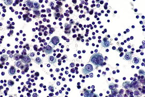
Reactive pleural effusion showing mesothelial cells lymphocytes neutrophils and macrophages. The fluid can accumulate quickly if the mesothelial cells fail to function resulting in an unhealthy collection of fluids in the chest cavity.
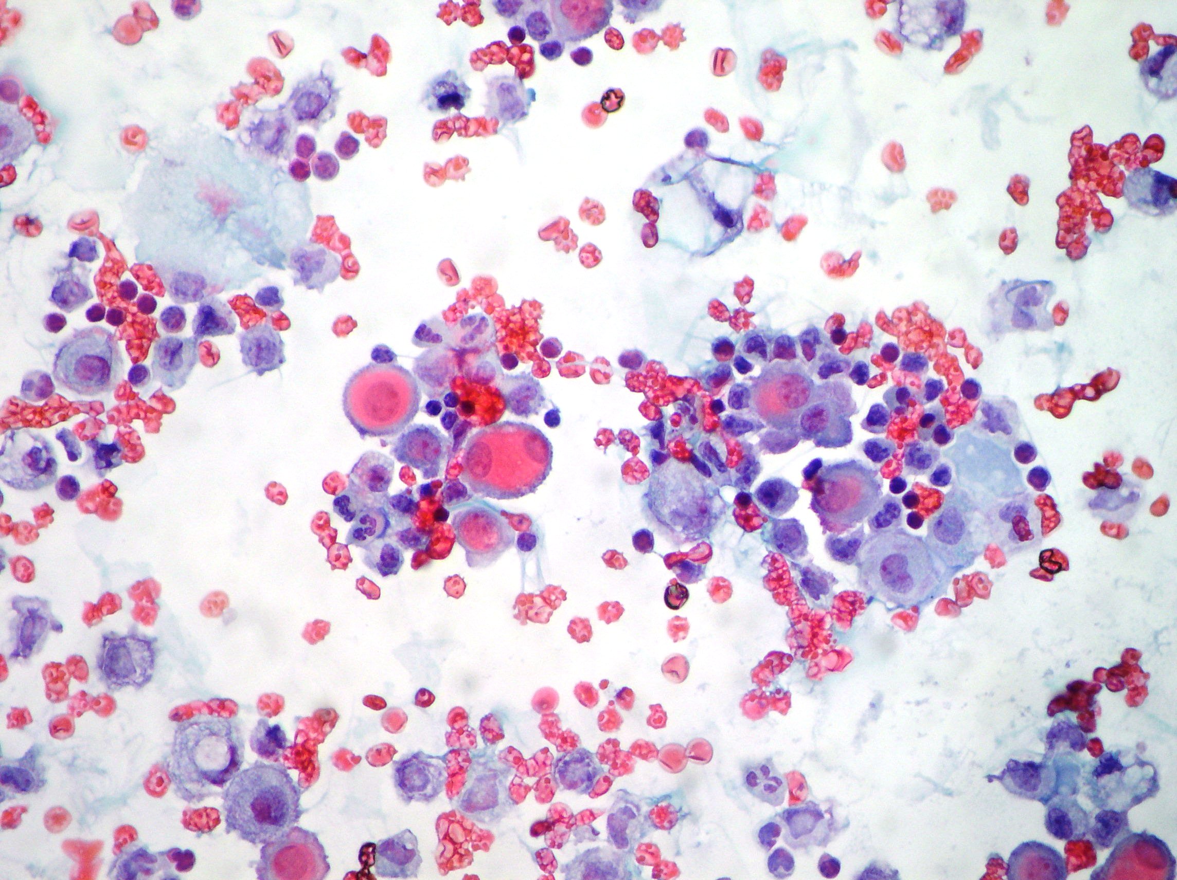


Effusions Cytopathology Cellnetpathology

Numerous reactive mesothelial cells were present in only 12 of specimens examined.
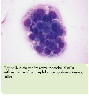
Mesothelial cells in pleural fluid. Reactive mesothelial cells present in a background of abundant lymphocytes. This has a large ddx. This condition can be due to the presence of a bacterial viral or fungal infection.

It can also be the result of trauma or the presence of metastatic tumor. Reactive mesothelial cells reactive mesothelial cells in pleural fluid reactive mesothelial cells are found when there is infection or inflammation present in a body cavity. Pleural effusion mesothelial cells pleural effusion.

Negative for malignant cells. Pleural fluid right thoracentesis. Neoplastic transformation of mesothelial cells results in malignant mesothelioma an aggressive tumor especially the pleura.

Immunocytochemistry test resulted positive for calretinin marker antibody papanicolaou x100 x200 mesothelial cells pleuric mesothelioma. Numerous mesothelial cells are seen in this pleural fluid from a dog with a transudative effusion with concurrent diapedesis of red blood cells or hemorrhage. Mesothelial cells are found in variable numbers in most effusions but their presence at greater than 5 of total nucleated cells makes a diagnosis of tb less likely.

This is known as pleural effusion. Reactive mesothelial cells can be found when there is an infection or an inflammatory response present in a body cavity. Actively dividing mesothelial cells can mimic an adenocarcinoma.

Pleural fluid for total white blood cell wbc count and differential cell count should be sent in an anticoagulated tube. A patient may develop shortness of breath and vague chest pains while the fluid experiences a buildup in the chest cavity these are two of the most common. Eighty five samples of pleural fluid obtained from 76 patients with biopsy proven tuberculous pleurisy were examined cytologically.

Specific diagnoses benign eosinophilic pleuritis general. Mesothelial cells in pleural fluid. Additional sampling should be considered within the clinical context.

The mesothelial cells have central round nuclei with a moderate amount of light purple cytoplasm and a corona or fringe to the cytoplasmic borders. In contrast 653 of pleural fluid aspirates obtained from a control group of pati. There are certain cells that line the pleura the thin double layered lining which covers the lungs chest wall and diaphragm which are known as mesothelial cellsother than the pleura mesothelial cells also form a lining around the heart pericardium and the internal surface of the abdomen peritoneum.

Use of pleural fluid n. Trauma with air in the pleural cavity.


Http Www Cap Org Apps Docs Committees Hematology Educational Activities 2009 Cmb Pdf
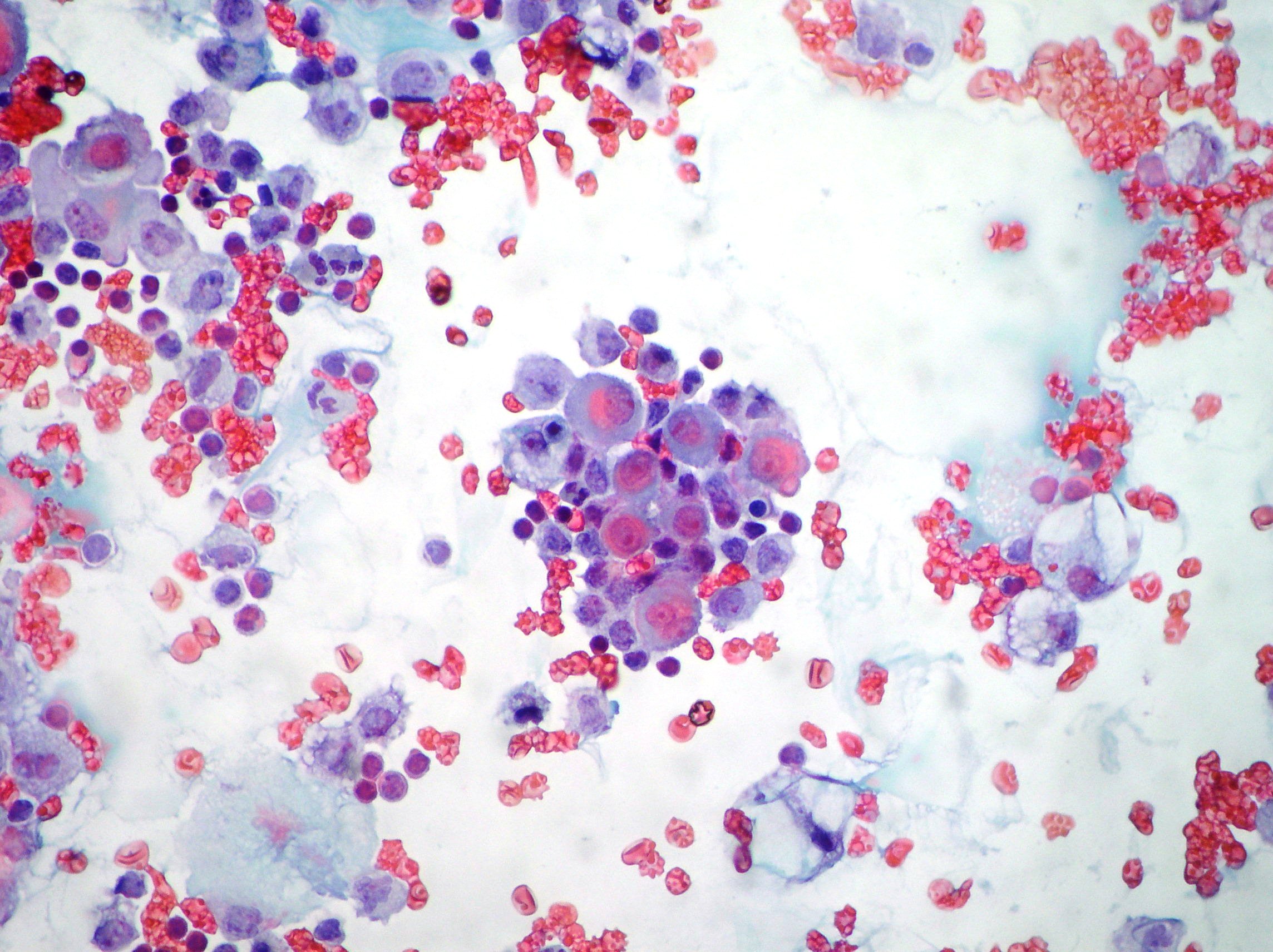


Effusions Cytopathology Cellnetpathology


Pleural Fluid Smear Malignant Mesothelial Cells Lymphocytes Mgg Download Scientific Diagram
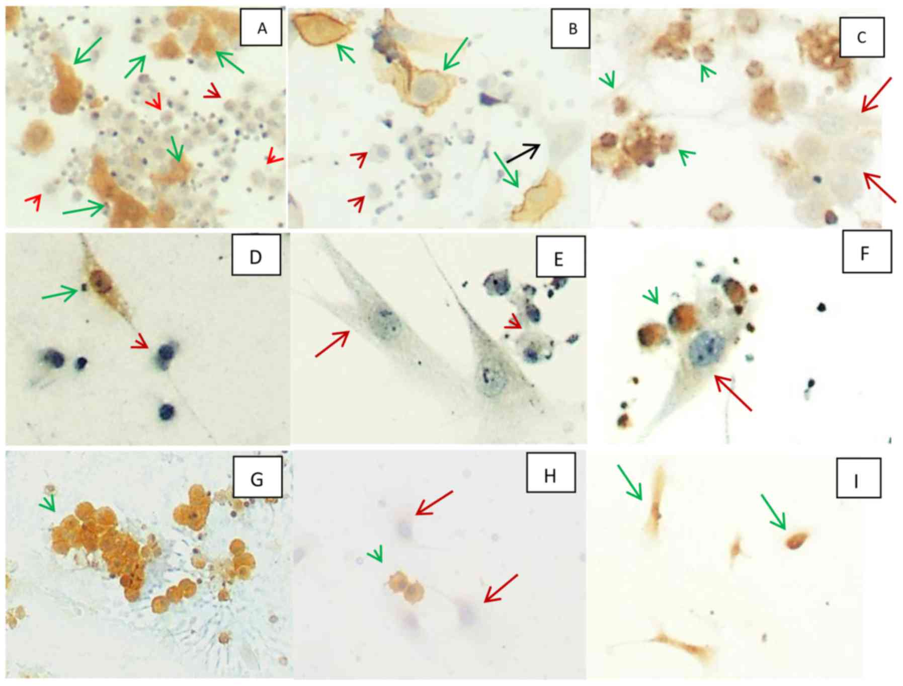


A Panel Of Markers For Identification Of Malignant And Non Malignant Cells In Culture From Effusions


View Image


Hjcam Iatrikh Zwwn Syntrofias Hellenic Journal Of Companion Animal Medicine Volume 6 Issue 1 2017 Pleural Effusion In The Cat A Focus On Laboratory Diagnosis


Http Www Cap Org Apps Docs Committees Hematology Educational Activities 2009 Cmb Pdf
Mesothelial Cells In Pleural Fluid Tuberculosis

Tuberculous pleurisy is the most frequent extrapulmonary manifestation of tuberculosis. 643 and 3 514.


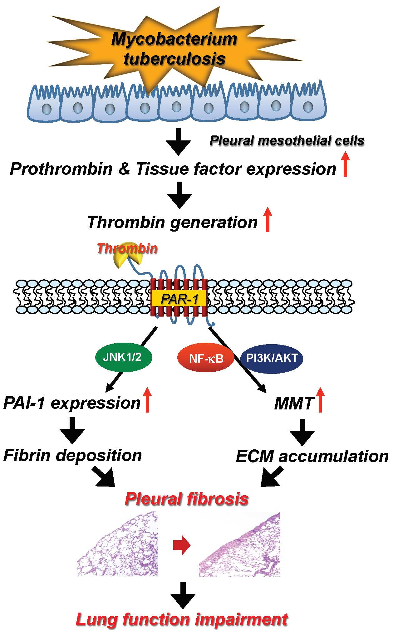
Ijms Free Full Text Thrombin Upregulates Pai 1 And Mesothelial Mesenchymal Transition Through Par 1 And Contributes To Tuberculous Pleural Fibrosis Html

The culture of the pleural fluid grew m tuberculosis.

Mesothelial cells in pleural fluid tuberculosis. 1 the pleura is a serous membrane that covers the lung parenchyma mediastinum diaphragm and rib cages and is divided into the visceral and parietal pleura. We report three hiv infected patients with tuberculous pleural effusions in which mesothelial cells were found in significant numbers in the pleural fluid. Numerous reactive mesothelial cells were present in only 12 of.

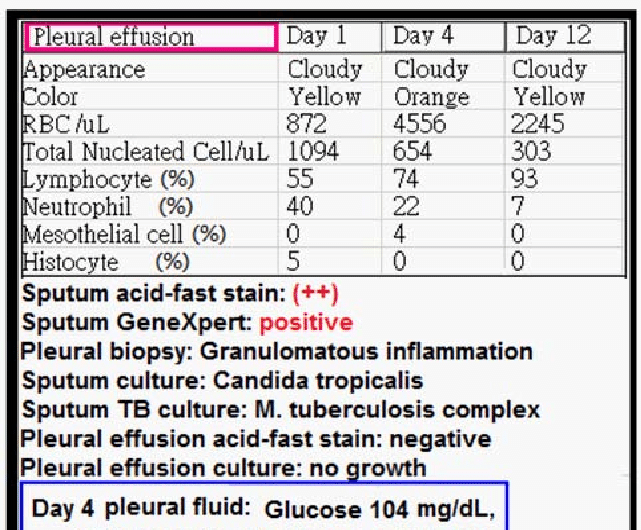
7 neutrophils 22 lymphocytes 60 macrophages and 10 mesothelial cells. The scarcity of mesothelial cells is a well known characteristic of tuberculous pleural effusions. Sensitivity of polymerase chain reaction to detect mycobacterium tuberculosis in pleural fluid varies from 40 to 80 percent and.

The patient was treated with a combination of six anti tb medications and was started on an antiretroviral regimen. Eighty five samples of pleural fluid obtained from 76 patients with biopsy proven tuberculous pleurisy were examined cytologically. Numerous reactive mesothelial cells were present in only 12 of specimens examined.

Pleural fluid cultures grow m tuberculosis in less than 65 of cases. Eighty five samples of pleural fluid obtained from 76 patients with biopsy proven tuberculous pleurisy were examined cytologically. Both the visceral and parietal pleurae are lined with a single layer of flat mesothelial cells that have some similarity of epithelial.

1 the fluid is generally an exudate 2 characterized by a predominance of lymphocytes and a paucity or absence of mesothelial cells3 4 5 in fact it has been concluded that the presence of numerous mesothelial cells almost excludes a diagnosis of tuberculosis. Pleural fluid from patients with tuberculosis had cells with patterns 1 914. The cytology showed reactive mesothelial cells and the differential cell count was as follows.

In contrast the combination of histology and culture of pleural tissue obtained by pleural biopsy. 625 and 1 416. Mesothelial cells in ascitic fluid mesothelial cells in ascitic fluid the associated tumor antigen 90k is known to possess properties similar.

Pleural effusion may occur at any stage of active tuberculosis. The suggestion that the presence of numerous often very reactive mesothelial cells in pleural aspirate makes the diagnosis of tuberculosis is unlikely confirmed mesothelial cells in pleural fluid. Mesothelial cells are found in variable numbers in most effusions but their presence at greater than 5 of total nucleated cells makes a diagnosis of tb less likely.

357 whereas most fluid secondary to cancer had cells with patterns 4 1016. This report concerns two cases of tuberculous pleural effusion in. Population 1 plus cells of variable size and complexity probably corresponding to macrophages and mesothelial cells in fluid.

In contrast 653 of pleural fluid aspirates obtained from a control group of pati. Actively dividing mesothelial cells can mimic an adenocarcinoma.


A Cytology Slide Demonstrating Numerous Mesothelial Cells In The Download Scientific Diagram


Https Encrypted Tbn0 Gstatic Com Images Q Tbn 3aand9gcravd78oblzuefp2w 9ddg Epzymjjo Zxiui9utmi09dnzwvdg Usqp Cau


Plos One Differentiation And Recruitment Of Th9 Cells Stimulated By Pleural Mesothelial Cells In Human Mycobacterium Tuberculosis Infection


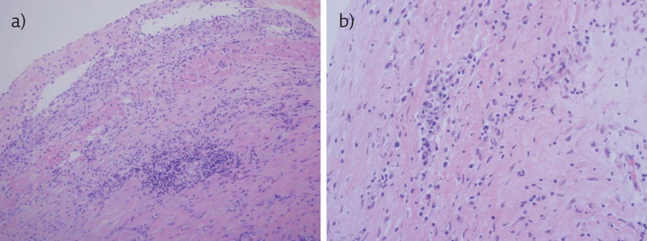
An Uncommon Cause Of Pleural Effusion European Respiratory Society


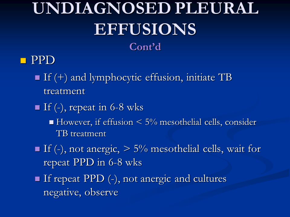
Approach To Pleural Effusion Ppt Download


Pleural Fluid Serial Analysis Reveals Lymphocytic Predominance Few Download Scientific Diagram


Tuberculous Pleural Effusion Shaw 2019 Respirology Wiley Online Library