Tag Archives: mesothelioma ct scan findings

Mesothelioma Ct Scan

The computer puts them together to make a 3 dimensional 3d image. Ct scan if doctors suspect mesothelioma in their patients they may recommend various diagnostic tools including medical imaging procedures.


Https Encrypted Tbn0 Gstatic Com Images Q Tbn 3aand9gcqm2wnxdnjwxjiarbr9ietjmckxbf5eolhcrkbcawdyim9zjp0n Usqp Cau

The computed tomography scan is an important tool in this process because it can provide highly detailed information about the disease type location and metastasis.

Mesothelioma ct scan. Ct scans can help physicians to determine the stage of your tumor. Ct scans are useful for diagnosing mesothelioma. A ct scan is a test that uses x rays and a computer to create detailed pictures of the inside of your body.

The image shows abnormal tissue which could be malignant tumors. Learn more about this process and other diagnosis issues from the illinois mesothelioma attorneys at shrader associates llp. This scan also can show if it has spread to lymph nodes or other organs.

It takes pictures from different angles. A new technique called ct perfusion can show if cancer cells are spreading in the bloodstream. Ct or cat stands for computed axial tomography.

Still many doctors say the ct scan is the best for the chest and abdomen which are where mesothelioma forms. You usually have a ct scan in the x ray radiology department as an. Ct scans also help doctors determine if treatment is working and the effect it has had on tumor number and size.

These scans also help stage cancer determining how much it has spread to other tissues. In short the best scientific evidence to date suggests that while ct scans are a useful tool in the work up of mesothelioma they cannot be relied upon to be fully accurate. Ultimately a biopsy is the only way to diagnose mesothelioma although the ct scan helps direct when and where to biopsy.

Ct scans are often used to help diagnose mesothelioma.


Diagnosis Of Malignant Pleural Mesothelioma Stanford Health Care


Pleural Effusion In Mesothelioma Ct Scan Stock Image C013 9667 Science Photo Library


The Role Of 18f Fdg Pet Ct Integrated Imaging In Distinguishing Malignant From Benign Pleural Effusion


Mesothelioma Lung Cancer Ct Scan Photograph By Du Cane Medical Imaging Ltd
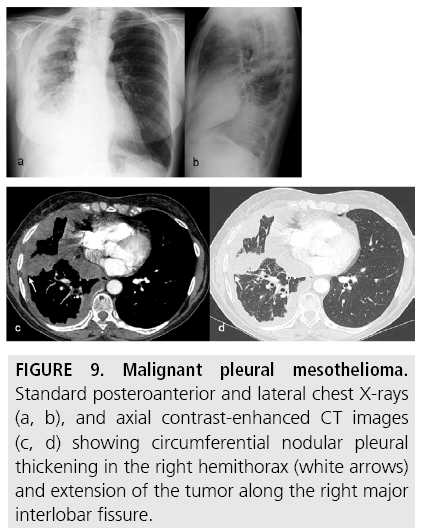


Diagnostic Imaging And Workup Of Malignant Pleural Mesothelioma


Pulmonary Radiologic Images


Metastatic Biphasic Pleural Mesothelioma Presenting With Cauda Equina Syndrome Sciencedirect
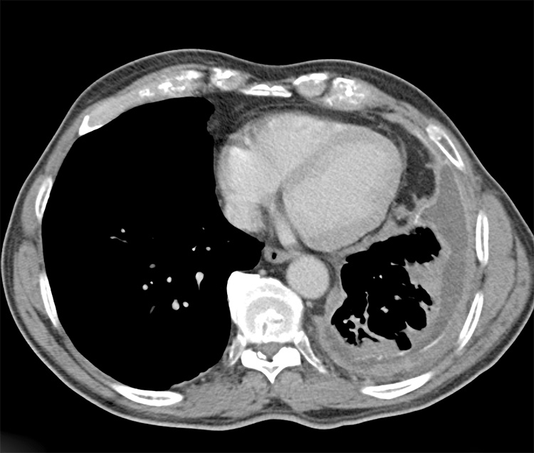
Mesothelioma Ct Scan Findings

Mesothelioma x ray findings ct scan and images if it is suspected that someone has mesothelioma or any other form of cancer a number of tests will be ordered. Ct is the primary imaging modality used for the evaluation of mpm.


Extrapleural And Chest Wall Extension From Malignant Mesothelioma In A Download Scientific Diagram

Given the presence of the mesothelium in different parts of the body mesothelioma can arise in various locations 17.

Mesothelioma ct scan findings. Mpm occurs more frequently in patients born or living in certain villages of turkeywe aimed to review radiological findings of mpmwe reviewed the ct findings in 219 biopsy proven mpm patients. There can be symptoms associated with hypercoagulability particularly due to a paraneoplastic syndrome associated with mesothelioma. Presenting with abdominal pain ct features of peritoneal masses and little to no ascites.

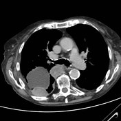
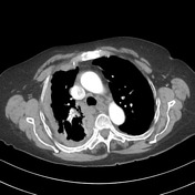
Here we show the ct scan of the chest from the same patient diagnosed with advanced mesothelioma. Common mri findings include. Computed tomography scan of a 58 year old patient with mesothelioma and shortness of breath.

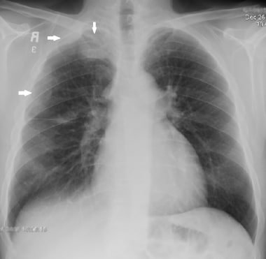
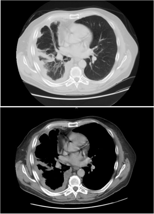
Learn more about this process and other diagnosis issues from the illinois mesothelioma attorneys at shrader associates llp. This image shows the extensive pleural thickening that is characteristic of mesothelioma effusion and reduction in the volume of the affected. Ct scan findings are similar to those of plain films but are seen better and in more detail.

Most tumors arise from the pleura and so this article will focus on pleural mesothelioma. Our team is working and offering consultations via phone e mail and video conferencing. Ct scans are often used to help diagnose mesothelioma.

Furthermore pleural thickening and effusion can be distinguished with ct scanning. Key ct findings that suggest mpm include unilateral pleural effusion fig 1 nodular pleural thickening figs 2 4 and interlobar fissure thickening fig 5. Many of the tests are non invasive and they help physicians to determine the cause of certain symptoms.

Malignant pleural mesothelioma mpm is an uncommon neoplasm. Solitary foci of chest wall invasion endothoracic fascial involvement and diaphragmatic invasion. Pleural mesothelioma 90 covered in this article.

Ct scan if doctors suspect mesothelioma in their patients they may recommend various diagnostic tools including medical imaging procedures. Ct scans and mri tests do not provide an unequivocal diagnosis of mesothelioma so tissue biopsy is required for definitive diagnosis. The computed tomography scan is an important tool in this process because it can provide highly detailed information about the disease type location and metastasis.

Growth typically leads to tumoral encasement of the lung with a rindlike appearance. Mesothelioma also known as malignant mesothelioma is an aggressive malignant tumor of the mesothelium. Diaphragmatic invasion ascites and omental caking are common ct scan findings of peritoneal mesothelioma.

There is a reported correlation between the clinical presentation and ct findings with subgrouping into 2.


Insert The Dark Side Of Mesothelioma Sciencedirect


A 71 Year Old Patient With Pleural Mesothelioma Ct Scan Top A Download Scientific Diagram


Study Intrapleural Air Flow Can Enhance Mesothelioma Ct Scans


Https Encrypted Tbn0 Gstatic Com Images Q Tbn 3aand9gcqwc6oe7ttcb Hourhci6giifatc6iektdkoon6yewe6pzkhm69 Usqp Cau


Mesothelioma Radiology Reference Article Radiopaedia Org


Mesothelioma Radiology Reference Article Radiopaedia Org


Malignant Mesothelioma Imaging Overview Radiography Computed Tomography