Tag Archives: mesothelioma cytology features

Mesothelioma Cytology Features

Regardless of the presence or absence of these. Tubular papillary microcystic solid adenomatoid decidual pleomorphic clear cell small cell.


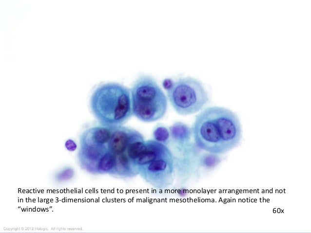
Malignant Mesothelioma In Effusions And Fine Needle Aspirates No Relationship Exists That Represents A Possible Conflict Of Interest With Respect To The Ppt Download

Overall when observing the cytology of malignant mesothelioma and other cancers researchers tend to focus on certain microscopic features of the structure and function of the cells.

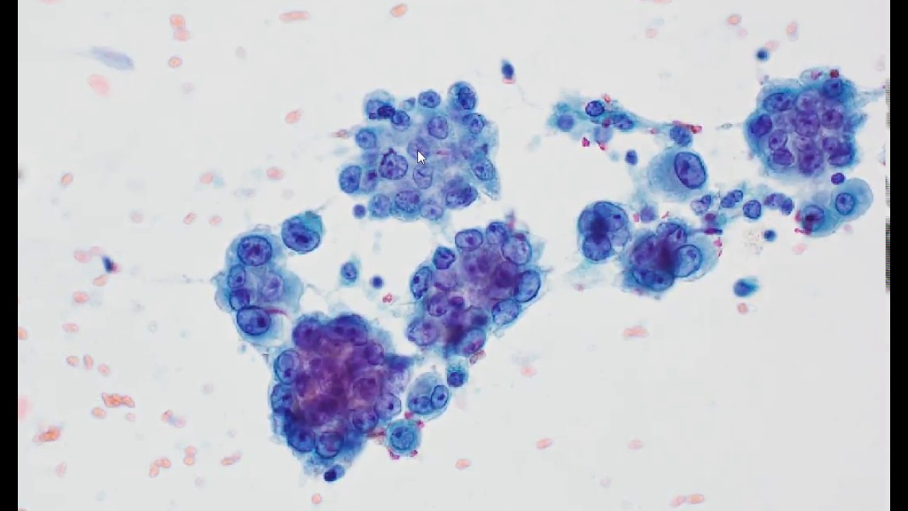
Mesothelioma cytology features. Large clusters with scalloped knobby edges. Pathologists can gather pleural fluid called a pleural effusion from a patient suspected of having pleural cancer. The diagnosis of malignant mesothelioma in effusion cytology a reappraisal and results of a multiinstitution survey.

It is taken from the pleural space outside of the lung using a fine. Cytologic appearance considered to be pathognomonic. Bap1 immunohistochemical ihc staining of malignant mesothelioma mm and reactive mesothelial cell rmc proliferations in cytology samples.

History of rheumatoid arthritis. The epithelioid type which is the most common can show different growth patterns. Cytology studies cells from fluids or surface scrapings without obtaining actual tissue samples.

May have epithelioid morphology. Necrotic debris fluffy orange to blue crap. Mesothelioma cytology or mesothelioma cytopathology is the study of cells for the presence of mesothelioma.

The diagnosis of mesothelioma requires a combination of appropriate clinical features past asbestos exposure persistent effusion consistent radiological findings and characteristic histology and cytology. It is a part of mesothelioma pathology which is the study of tissue or fluid to determine if this cancer exists. All mesothelioma biopsycytology pairs showed the same pattern of bap1 or p16 retention or loss in the biopsy and cytology specimens am j surg pathol 201640120126 figure 1.

A desmoplastic variant of mesothelioma is also described. Large single multinucleated cells classically spindled. With a better understanding of these and other characteristics researchers may ultimately be able to decipher how the cancer may spread and react to treatment.

Cytologic features cytology pleural mesothelioma fluid stain. 2 key features of mesothelioma include complex architectural arrangements necrosis and an expansile and disorganized growth pattern.


Diagnostic Challenge Department Of Pathology Pdf Free Download


Effusion Cytology Clinician S Brief


Https Encrypted Tbn0 Gstatic Com Images Q Tbn 3aand9gcsbgvpsjkfmxbyb2dtepoaktmrusji5uunvmadtoap8gksrx8cp Usqp Cau


What S New In Mesothelioma Pathologica Journal Of The Italian Society Of Anatomic Pathology And Diagnostic Cytopathology


A Cytological Features Of Malignant Mesothelioma Papanicolaou Download Scientific Diagram


A Cytological Features Of Malignant Mesothelioma Papanicolaou Download Scientific Diagram
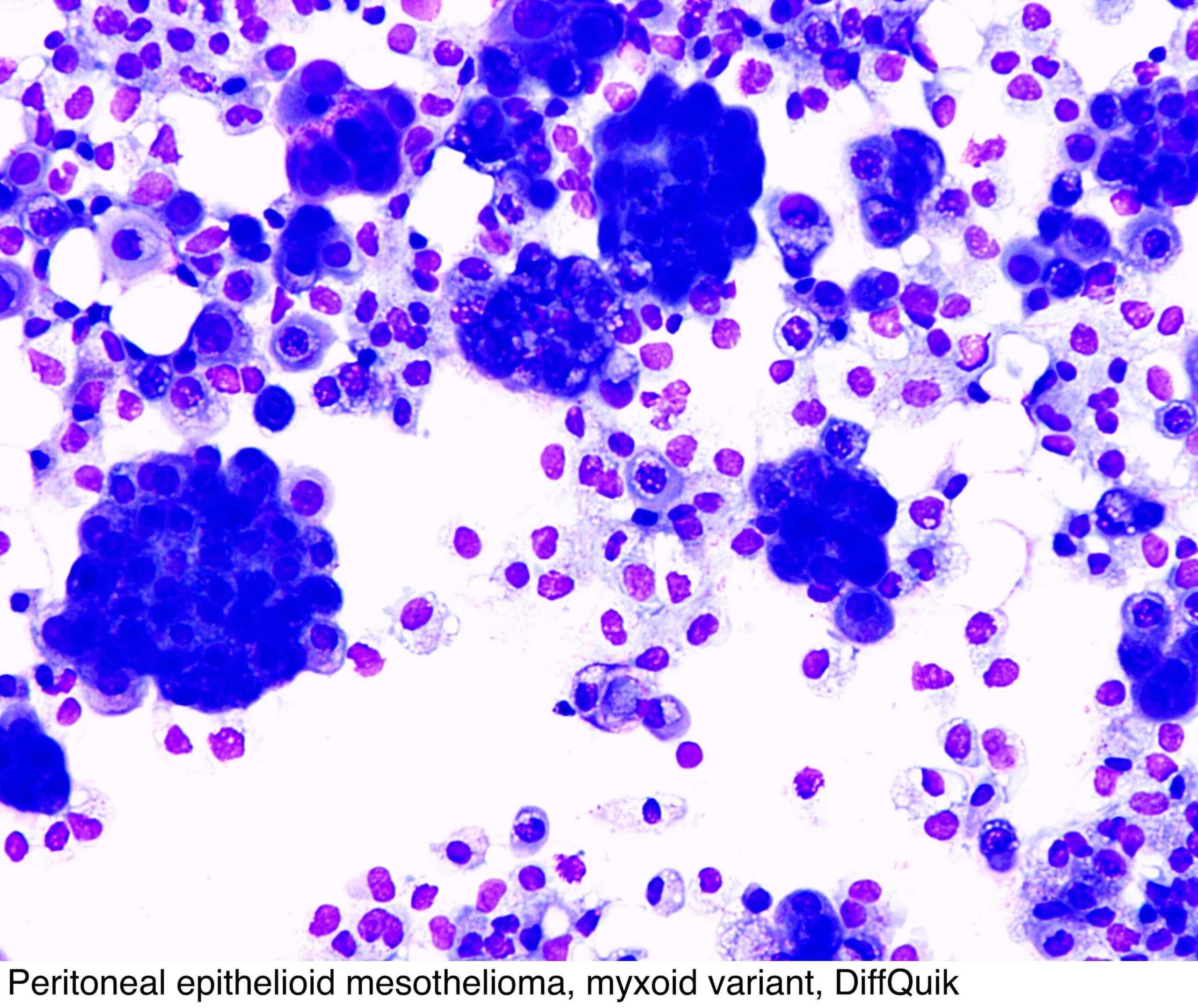


Fluid Cytology In Serous Cavity Effusions

Mesothelioma Cytology

It deals with pericardial fluid peritoneal fluid and pleural fluid. Signs and symptoms of mesothelioma may.


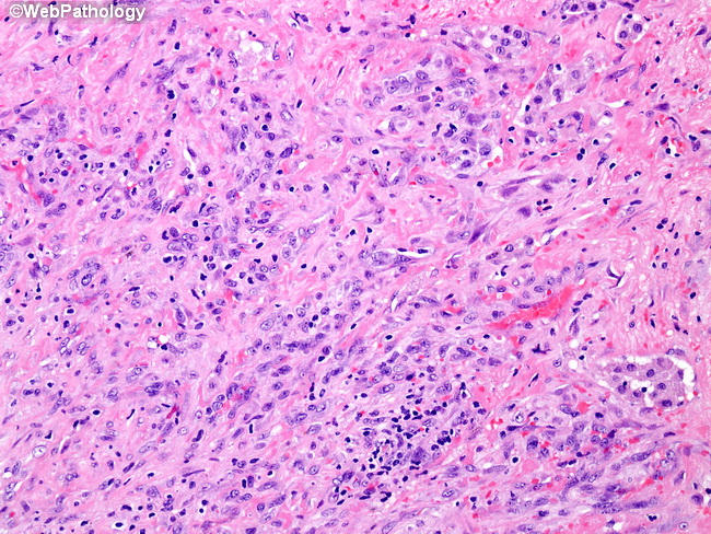
Webpathology Com A Collection Of Surgical Pathology Images

This fact is due to tissue samples being more reliable than fluid samples.

Mesothelioma cytology. It is a part of mesothelioma pathology which is the study of tissue or fluid to determine if this cancer exists. One study published on uptodate compared the accuracy of tissue biopsies and fluid samples. Mesothelioma cytology unlike imaging tests cytology is a pathological means to diagnosing malignant mesothelioma in patients consisting of a fluid biopsy of abnormalities previously discovered.

Mesothelioma cytology or cytopathology goes hand in hand with histology. However there is still a small risk of tumor cell seeding microscopic spreading of cancer due to cells moving during a biopsy during cytologic procedures. A look at how the cancer will advance.

Mesothelioma histology is more trusted than cytology for pathologically diagnosing this disease. By forgoing a tissue biopsy there is less risk of morbidity of the patient. Less commonly the lining of the abdomen and rarely the sac surrounding the heart or the sac surrounding the testis may be affected.

The article deals with cytopathology specimens from spaces lined with mesothelium ie. The most common area affected is the lining of the lungs and chest wall. While the histological determination will focus more on the cell type the cytology research will dive deeper into how these cells are forming interacting and spreading in the body.

Mesothelial cytopathology is a large part of cytopathology. Mesothelioma cytology or mesothelioma cytopathology is the study of cells for the presence of mesothelioma. Mesothelioma is a type of cancer that develops from the thin layer of tissue that covers many of the internal organs known as the mesothelium.

Cytology is often one of the first tests used in the diagnosis of mesothelioma after imaging tests reveal the possibility of cancer. Mesothelioma cytology is a useful tool that enables analysis without necessitating a tissue biopsy. Carlos wm bedrossian an update on pleuropulmonary cytopathology.

Once cytology is performed the cells from the sample are examined to arrive at a diagnosis. Cytology or fluid biopsy is a pathology specialty which studies disease by looking at the appearance and behavior of cells in bodily fluids. Mesothelioma cytology mesothelioma cytology.

Cytological diagnosis of mesothelioma and molecular cytology of lung cancer with an historical perspective diagnostic cytopathology 101002dc23298 43 7 513 526 2015.


Figure 2 A B Neoplastic Cells Arrows Intermixed With Reactive Mesothelial Cells Note The Eccentric Location Of The Nuclei Of Neoplastic Cells Kletki Krovi


Moving Toward An Earlier Diagnosis Of Mesothelioma


Cytological Profile Of A Case Of Malignant Mesothelioma Of Pleura With Supraclavicular Lymph Node Metastasis Abstract Europe Pmc


Https Onlinelibrary Wiley Com Doi Pdf 10 1002 Cncy 21342


Mesothelioma Dog


Https Encrypted Tbn0 Gstatic Com Images Q Tbn 3aand9gcsbgvpsjkfmxbyb2dtepoaktmrusji5uunvmadtoap8gksrx8cp Usqp Cau


The Value Of Brca 1 Associated Protein 1 Expression And Cyclin Dependent Kinase Inhibitor 2a Deletion To Distinguish Peritoneal Malignant Mesothelioma From Peritoneal Location Of Carcinoma In Effusion Cytology Specimens Althakfi 2020