Tag Archives: mesothelioma histology microvilli

Mesothelioma Histology Microvilli

We studied the reactivity of malignant mesothelioma cells with tumor markers and the phenotypes of lymphocyte subsets in pleural effusions from 14 patients with malignant mesothelioma. The mesothelium is a membrane composed of simple squamous epithelium that forms the lining of several body cavities.
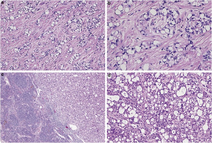


Mesothelioma With Signet Ring Cell Features Report Of 23 Cases Modern Pathology

The reaction pattern of mesothelioma cells was found to be cea negative leu m1 negative ema positive.

Mesothelioma histology microvilli. Mesothelial tissue also surrounds the male internal reproductive organs the tunica vaginalis testis and covers the internal reproductive organs of. Mesothelioma histology involves the study of cancerous mesothelial cells. Histology technicians make use of microscopes to visualize tissue and cells accurately.

Doctors use these histological classifications to confirm the diagnosis. It is a branch of histopathology which studies diseased cells. With a distinctly round or cuboidal shape and elongated microvilli when viewed under a microscope.

Mesothelioma tumors typically present with three distinct histological abnormalities. Mesothelioma histology is the study of mesothelial cancer cells. Histology is an extended branch of biology in which cells and tissues are studied.

Mesothelioma histology an important tool used in the definitive diagnosis of disease is histology the microscopic examination of cellular anatomy. For identification of cell surface antigens with monoclonal antibodies the adhesive slide assay was used. Based on nine cases of mesotheliomas the sensitivity for long microvilli as a marker for mesothelioma is 889 and the specificity is 100.

Significance of studying mesothelium histology the study of histology of unnatural or diseased cells is termed histopathology. A fine needle aspiration biopsy of a pleural lesion resulted in a fragment of tissue composed of loosely aggregated tumor cells many of which have a considerable portion of the cell surface covered with microvilli m. The pleura thoracic cavity peritoneum abdominal cavity including the mesentery mediastinum and pericardium heart sac.

Histopathology of mesothelioma cells provide various insight to the illness and is a methods of accurate diagnosis. Histopathological studies can only be conducted with a certified and experienced medical professional. Hammar et al 21 reported four cases of mucicarmine positive mesothelioma in which tubular crystalloids were found to be associated with the microvilli in all four cases as well as in the.

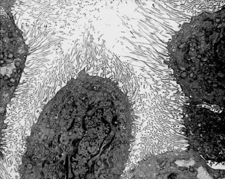
The microvilli are profuse and rather than appearing to arise individually as straight projections. Epithelial cells have microvilli microscopic protrusions of the cell and clear structures called organelles within each cell. Long microvilli were observed in eight of nine cases of mesothelioma but not in 50 cases of mesothelial hyperplasia or 50 cases of nonmucinous pulmonary adenocarcinoma fig.


Malignant Mesothelioma Of Pleura Thoracic Pathology A Volume In The High Yield Pathology Series Expert Consult Online And Print 1st Edition
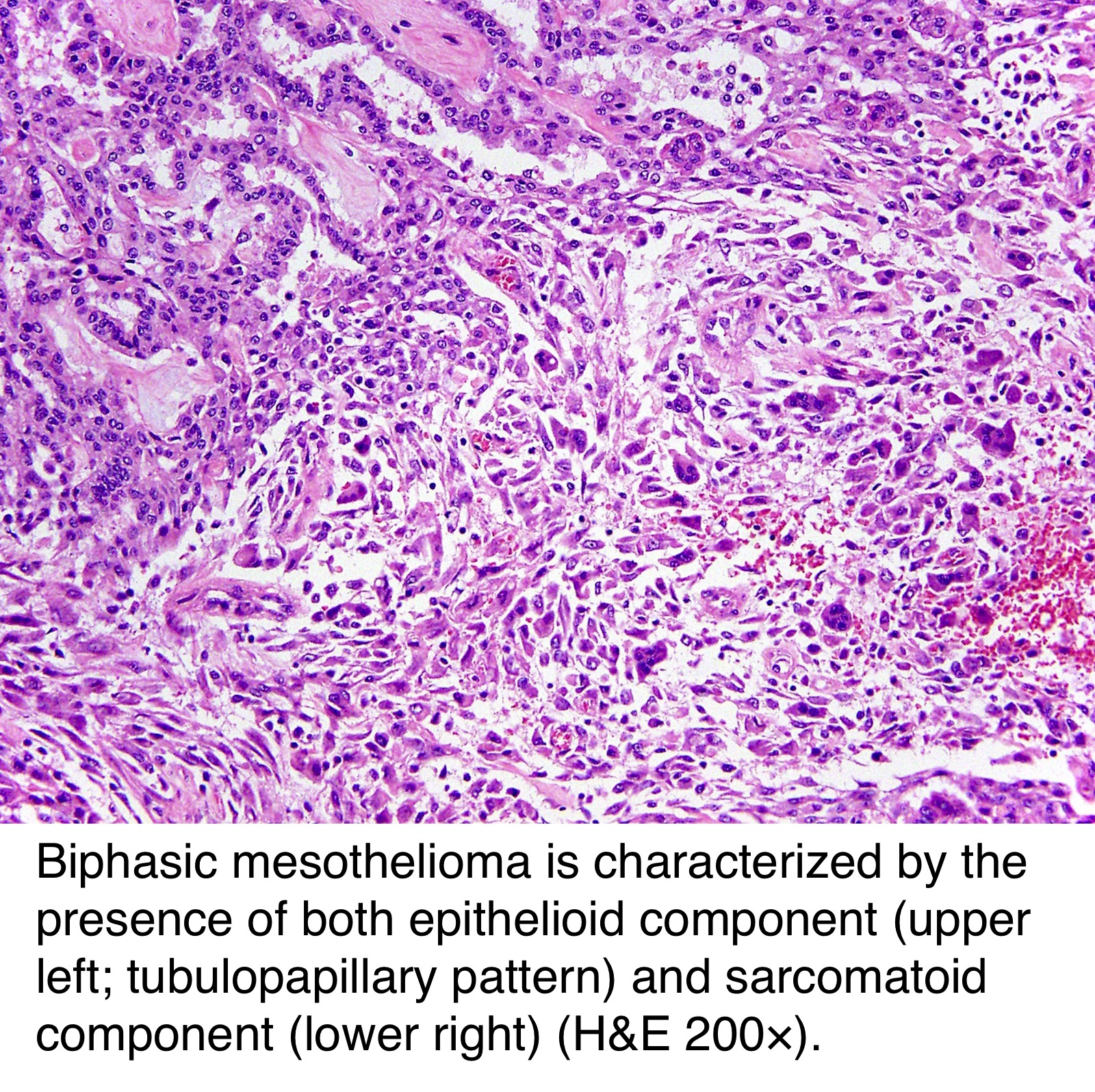


Pathology Outlines Peritoneal Malignant Mesothelioma
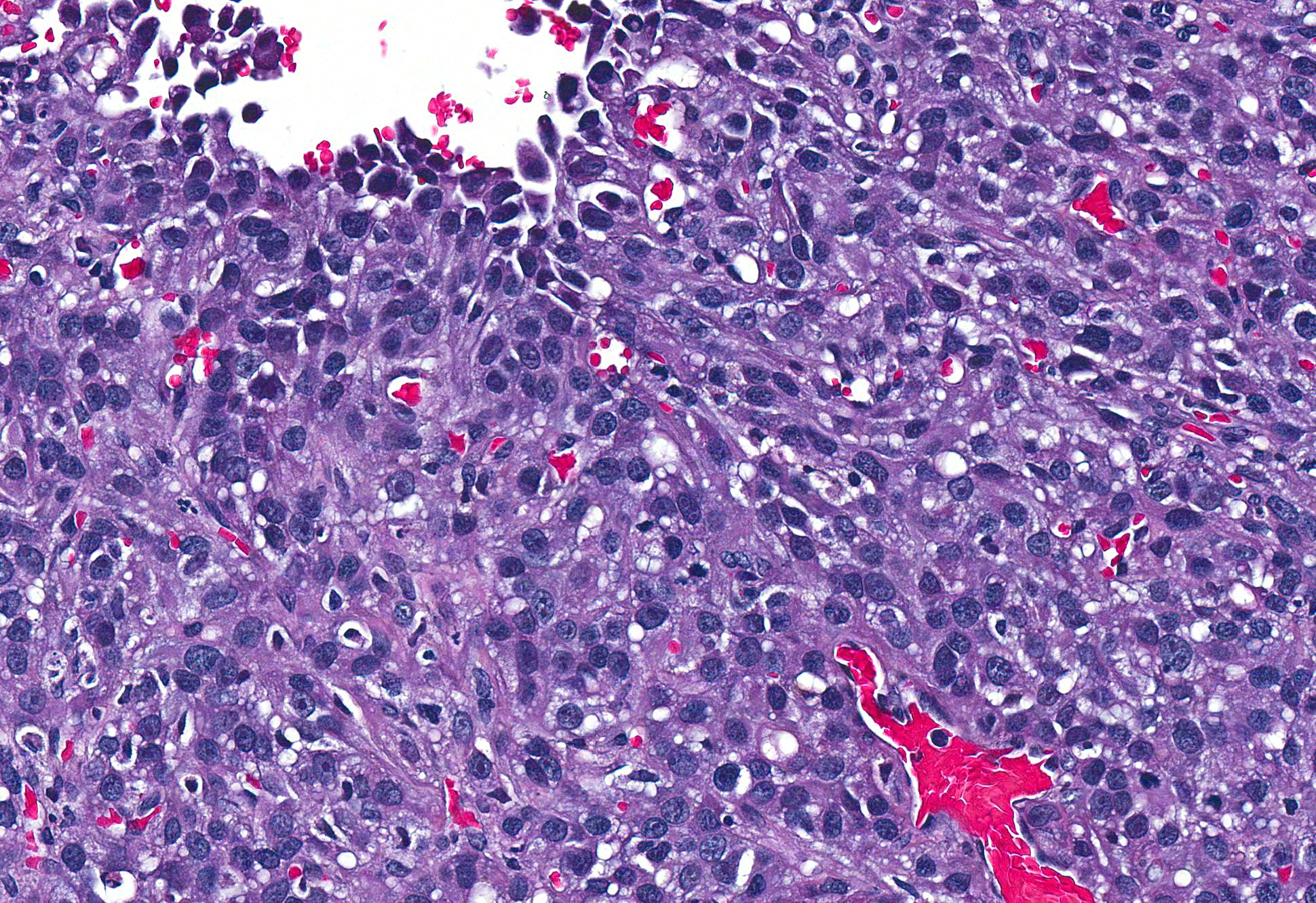


Conference 7 2017 Case 2 20171018


Early Detection Of Malignant Pleural Mesothelioma


Pdf Sarcomatoid Mesothelioma A Clinical Pathologic Correlation Of 326 Cases


Https Encrypted Tbn0 Gstatic Com Images Q Tbn 3aand9gcs9q5qyx6e27peswq P1qgcnejk5dfbyccejazpqk2e2yl7mico Usqp Cau


Http Eknygos Lsmuni Lt Springer 266 Part 20six 6 20item Pdf

Mesothelioma Histology

Histopathology falls within the larger field of pathology. Mesothelioma histology involves the study of cancerous mesothelial cells.


Primary Peritoneal Epithelioid Mesothelioma Of Clear Cell Type With A Novel Vhl Gene Mutation A Case Report Sciencedirect

Mesothelioma is a type of cancer that develops from the thin layer of tissue that covers many of the internal organs known as the mesothelium.

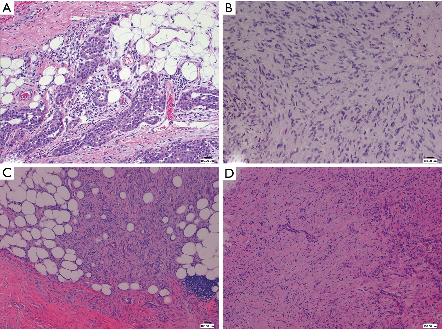
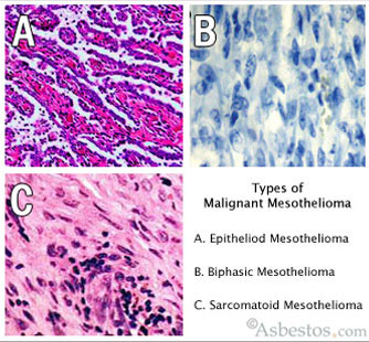
Mesothelioma histology. Observing the anatomy of a particular cancer or how the tumors grow and develop is one way of diagnosing mesothelioma. This form of mesothelioma is comprised of cells which resemble the normal mesothelial cells in that they are arranged in a trabecular fashion. Pathology including histopathology and cytology analyses helps doctors determine the mesothelioma cell type stage and how the cancer is expected to progress.

Mesothelioma histology involves two steps. Mesothelioma pathology is an important aspect of reaching an accurate diagnosis. Histology is a branch of biology that involves the study of cells and tissues.

The most common area affected is the lining of the lungs and chest wall. The tissue samples are sent to a laboratory where an experienced pathologist will review the samples and look for epithelioid sarcomatoid or biphasic mesothelioma cells. This process is part of mesothelioma pathology which involves examining either tissue or fluid to determine if this cancer exists in the body.

The pathology of tumor growth. Mesothelioma pathology provides a full picture of the cancer contributing to a more accurate diagnosis and an informed treatment plan. Well differentiated papillary mesothelioma.

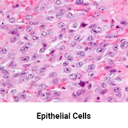
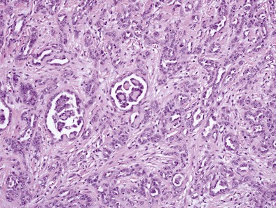
Histologically epithelial mesothelioma cells have polygonal ovoid or cuboidal cell shape. Signs and symptoms of mesothelioma may. The occasional presence of signet ring cells may make it challenging to distinguish this disease from cancers of the lung.

Doctors perform a tissue biopsy on a patient to collect tissue samples. The most obvious anatomical sign of mesothelioma is the. Papillae with myxoid cores each lined by a single mesothelial cell layer invasion is typically not present am j surg pathol 201438990 overall more indolent than peritoneal malignant mesothelioma ann surg oncol 201926852.

Most commonly in the peritoneum rarely pleura and other sites histology. Mesothelioma histology or mesothelioma histopathology is the study of tissue for the presence of mesothelioma. Less commonly the lining of the abdomen and rarely the sac surrounding the heart or the sac surrounding the testis may be affected.

Another subset of this field called histopathology looks specifically at the microstructure of diseased cells and tissues which is what a pathologist will focus on when studying a tissue sample to make a diagnosis. Mesothelioma histology is the study of the function and structure of anatomy including tissues and cells. Histopathology is the study of diseased cells.


Localized Malignant Pleural Mesothelioma Report Of Two Cases Journal Of Thoracic Oncology


Figure 3 From Pathology Of Mesothelioma Semantic Scholar


Malignant Pleural Mesothelioma Clinical Gate


The Pathological And Molecular Diagnosis Of Malignant Pleural Mesothelioma A Literature Review Ali Journal Of Thoracic Disease


Figure 4 From Pathology Of Mesothelioma Semantic Scholar


Challenges And Controversies In The Diagnosis Of Malignant Mesothelioma Part 2 Malignant Mesothelioma Subtypes Pleural Synovial Sarcoma Molecular And Prognostic Aspects Of Mesothelioma Bap1 Aquaporin 1 And Microrna Journal Of Clinical Pathology


Mesothelioma Asbestos Images Diagrams Graphs

Mesothelioma Microvilli

Based on nine cases of mesotheliomas the sensitivity for long microvilli as a marker for mesothelioma is 889 and the specificity is 100. Tem ultrastructure of cultured mesothelioma cells showed microvilli of various lengths in different cell lines but overall the microvilli in cultured mesothelioma cells were shorter than those observed in histological samples of mesothelioma.


Mesothelioma Microvilli

A fine needle aspiration biopsy of a pleural lesion resulted in a fragment of tissue composed of loosely aggregated tumor cells many of which have a considerable portion of the cell surface covered with microvilli m.

Mesothelioma microvilli. Mustaersataariuwaeduau mesothelial cells form a monolayer of specialised pavement like cells that line the bodys serous cavities and internal organs. 1department of surgery university of western australia royal perth hospital perth 6000 wa australia. Epithelial cells have microvilli microscopic protrusions of the cell and clear structures called organelles within each cell.

The luminal surface is covered with microvilli. Mesothelioma pleural mass effusion biopsy shows malignancy em shows long microvilli hx of asbestos exposure sob nonpleuritic chest pain hx of asbestos exposure. Long microvilli were observed in eight of nine cases of mesothelioma but not in 50 cases of mesothelial hyperplasia or 50 cases of nonmucinous pulmonary adenocarcinoma fig.

Hammar et al 21 reported four cases of mucicarmine positive mesothelioma in which tubular crystalloids were found to be associated with the microvilli in all four cases as well as in the. Mesothelioma histology involves the study of cancerous mesothelial cells. The mesothelium is composed of an extensive monolayer of specialized cells mesothelial cells that line the bodys serous cavities and internal organs.

Doctors use these histological classifications to confirm the diagnosis. The proteins and serosal fluid trapped by the microvilli provide a slippery surface for internal organs to slide past one another. Int j biochem cell biol.

Microvilli are characteristic of mesothelial cells.


Pdf Disseminated Lipid Rich Peritoneal Mesothelioma In A Horse


Length Dependent Retention Of Fibres In The Pleural Space Chapter 5 The Toxicology Of Carbon Nanotubes


Http Eknygos Lsmuni Lt Springer 266 Part 20six 6 20item Pdf


Wo2017014337a1 Method For Preparing Sample For Scanning Electron Microscopic Observation Of Pathogen Species Including Novel Paraffin Tissue For Diagnosis Of Malignant Mesothelioma Google Patents


Https Www Thoracic Theclinics Com Article S1547 4127 04 00122 7 Pdf


1


2016 Evening Specialty Conference Gynecologic Pathology

Mesothelioma Histo

Malignant mesothelioma me zoe thee lee o muh is a type of cancer that occurs in the thin layer of tissue that covers the majority of your internal organs mesothelium. It most commonly occurs in the linings of the lungs or the abdomen.


Pathology Outlines Diffuse Malignant Mesothelioma

Less commonly the lining of the abdomen and rarely the sac surrounding the heart or the sac surrounding the testis may be affected.

Mesothelioma histo. Testa jr cheung m pei j et al. The history of mesothelioma includes its discovery its association with asbestos and the regulation of asbestos use in workplaces schools buildings and ships. Histopathology is the study of diseased cells.

Symptoms can take 20 50 years to develop and become noticeable making for a poor prognosis. Histopathology falls within the larger field of pathology. Mesothelioma and asbestos history coincide because the use of the toxic mineral is strongly associated with the cancer.

Despite decades of research there still is no definitive cure for the four types of mesotheliomahowever more precise surgical procedures improved chemotherapy regimens and emerging treatments such as immunotherapy are helping patients survive longer than ever before. Mesothelioma treatments are available but for many people with mesothelioma a cure isnt possible. Signs and symptoms of mesothelioma may.

The most common area affected is the lining of the lungs and chest wall. The average life expectancy is 12 21 months after diagnosis but prognosis may improve with treatment. Family history of mesothelioma and other cancers including uveal melanoma renal cell carcinoma and cutaneous melanoma may suggest an underlying germline bap1 mutation.

Germline bap1 mutations predispose to malignant mesothelioma. Mesothelioma histology involves the study of cancerous mesothelial cells. History of mesothelioma treatment.

Mesothelioma is a type of cancer that develops from the thin layer of tissue that covers many of the internal organs known as the mesothelium. Mesothelioma is an aggressive and deadly form of cancer. Histology is a branch of biology that involves the study of cells and tissues.

A brief history of mesothelioma. This dangerous form of cancer affects 2000. Mesothelioma is a rare and aggressive form of cancer found in the protective lining mesothelium of the lungs chest heart abdomen and testicles.

Mesothelioma is a cancer caused by asbestos.


Pleura Revisited From Histology And Pathophysiology To Pathology And Molecular Biology Karpathiou 2019 The Clinical Respiratory Journal Wiley Online Library


Mesothelioma Cytology Youtube


Genetic Alterations Of Malignant Pleural Mesothelioma Association With Tumor Heterogeneity And Overall Survival Quetel 2020 Molecular Oncology Wiley Online Library


Https Www Researchgate Net Profile Richard Attanoos Publication 318287816 Guidelines For Pathologic Diagnosis Of Malignant Mesothelioma 2017 Update Of The Consensus Statement From The International Mesothelioma Interest Group Links 5adb02ca458515c60f5cd158 Guidelines For Pathologic Diagnosis Of Malignant Mesothelioma 2017 Update Of The Consensus Statement From The International Mesothelioma Interest Group Pdf


Antioxidants Free Full Text Mitochondrial Ros Induce Partial Dedifferentiation Of Human Mesothelioma Via Upregulation Of Nanog Html


Pdf Dissecting Heterogeneity In Malignant Pleural Mesothelioma Through Histo Molecular Gradients For Clinical Applications Semantic Scholar


Mesothelioma Histology Tonofilaments