Sarcomatoid mesothelioma like all other forms of the disease is caused by exposure to asbestos. Spindle cell neoplasms of the lung.

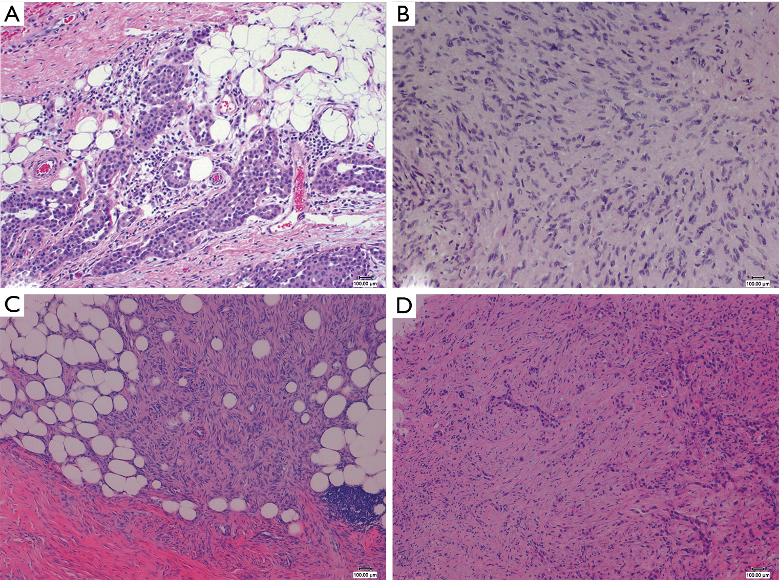
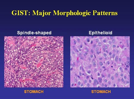
Under a microscope the cells of sarcomatoid mesothelioma are long and spindle shaped.
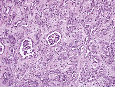
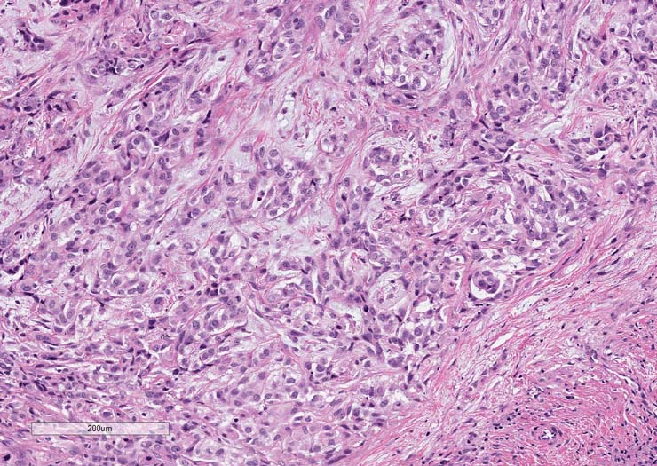
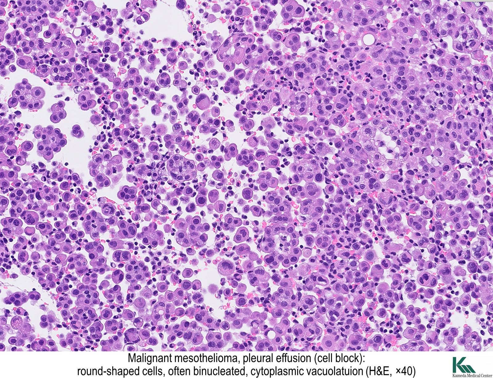
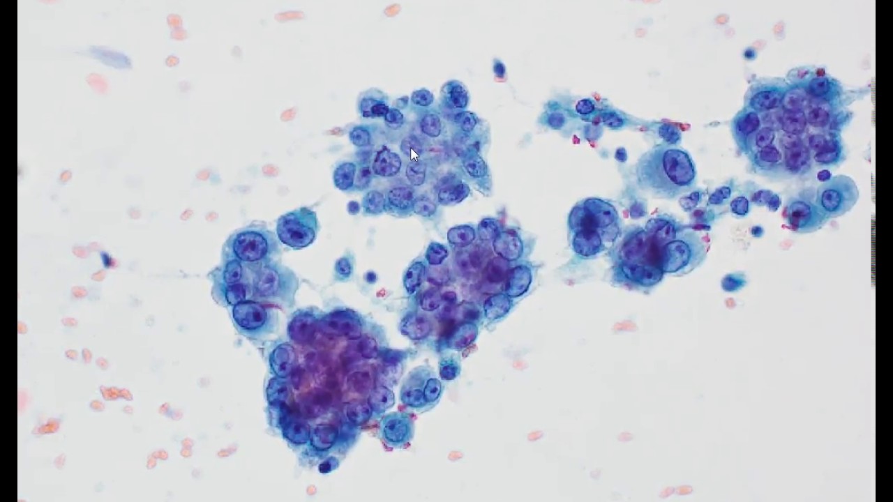
Mesothelioma spindle cell. Sarcomatoid mesothelioma is a malignant cell type of mesothelioma that is caused by asbestos exposure. Spindle cell tumors that arise in or metastasize to the pleura must be thoroughly evaluated to arrive at a definitive diagnosis. Sometimes have multiple nuclei.
Evidence based guidelines from the international mesothelioma panel and the mesopath national reference center hum pathol. Sarcomatoid mesothelioma is a rare cell type caused by asbestos exposure. Sarcomatoid cells however.
It accounts for approximately 10 20 of all mesothelioma diagnoses. It is estimated that 20 of mesothelioma cases are of this type. The cells are oval shaped much like a spindle and contain no nucleus.
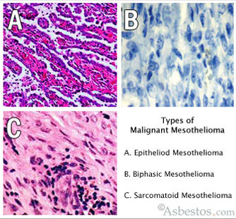
Spindle cell mesothelioma refers to a type of disease called sarcomatoid mesothelioma. Sarcomatoid carcinoma also referred to as spindle cell carcinoma or carcinosarcoma is a neoplasm of epithelial derivation that shows variable types of differentiation. Sarcomatoid mesothelioma is the least common and is often referred to as cell type or spindle cell mesothelioma.
Mesothelioma is rare cancer with only 3000 cases diagnosed annually but the prognosis is poor. The differential diagnosis between pleural sarcomatoid mesothelioma and spindle cellpleomorphic sarcomatoid carcinomas of the lung. Spread apart quick to metastasize.
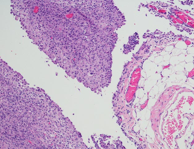
The cells are less uniform in structure and have a far more random arrangement than their epithelioid counterparts. Additionally many tumors arising in the lung and surrounding tissues involve the pleura. Malignant mesothelioma is the most common tumor arising in the pleura but metastatic tumors to the pleura occur more frequently.
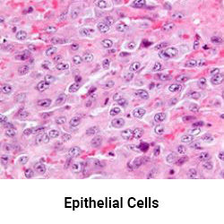
Sarcomatoid mesothelioma accounts for 10 20 of all cases and is the most aggressive but least common mesothelioma cell type. When comparing the malignant sarcomatoid cell type to malignant epithelial cells the most common cell type of mesothelioma epithelial cells are typically found clumped together in uniform formations. They appear similar to other types of cancer.
Primary pulmonary spindle cell neoplasms are uncommon and can occasionally be a source of diagnostic difficulty. The median survival for sarcomatous mesothelioma patients is typically less than 6 months. The spindle shaped sarcomatoid mesothelioma cells are rare and more aggressive than other mesothelioma cell types and metastasize quickly.
Sometimes referred to as spindle cell mesothelioma sarcomatoid cells are recognized by their oval spindle shape.
Https Patologi Com Guideline 20mesotheliom Pdf

Cd 26 Expression In Mesothelioma Cell Line Mesothelioma Cell Line Download Scientific Diagram
Sarcomatoid Mesothelioma Get Diagnosed Treated

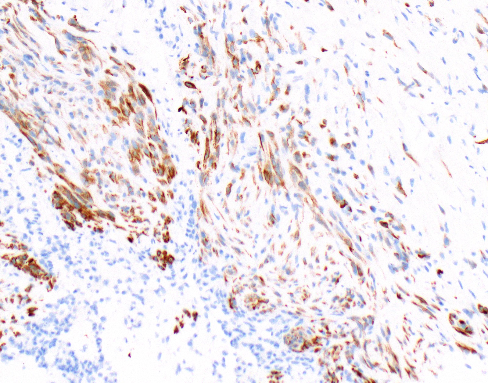
A Overview Of Spindle Cell Tumor With Densely Allocated Spindle Cells Download Scientific Diagram

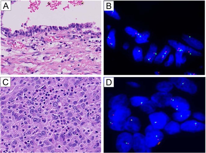
Pdf P1 09 003 Malignant Mesothelioma Versus Synovial Sarcoma An Analysis Of 19 Cases With Molecular Diagnosis
Pathology Outlines Diffuse Malignant Mesothelioma

Value Of Immunohistochemistry In The Differential Diagnosis Of Pleural Sarcomatoid Mesothelioma From Lung Sarcomatoid Carcinoma Semantic Scholar