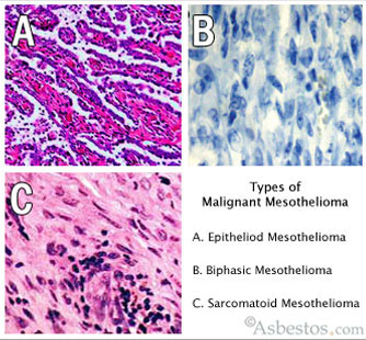
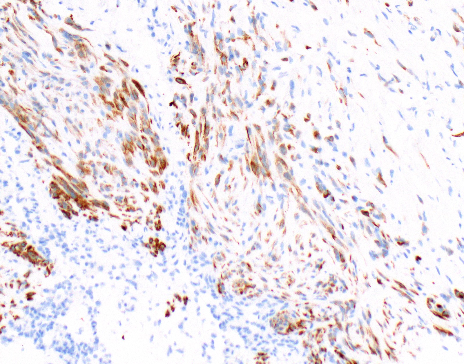
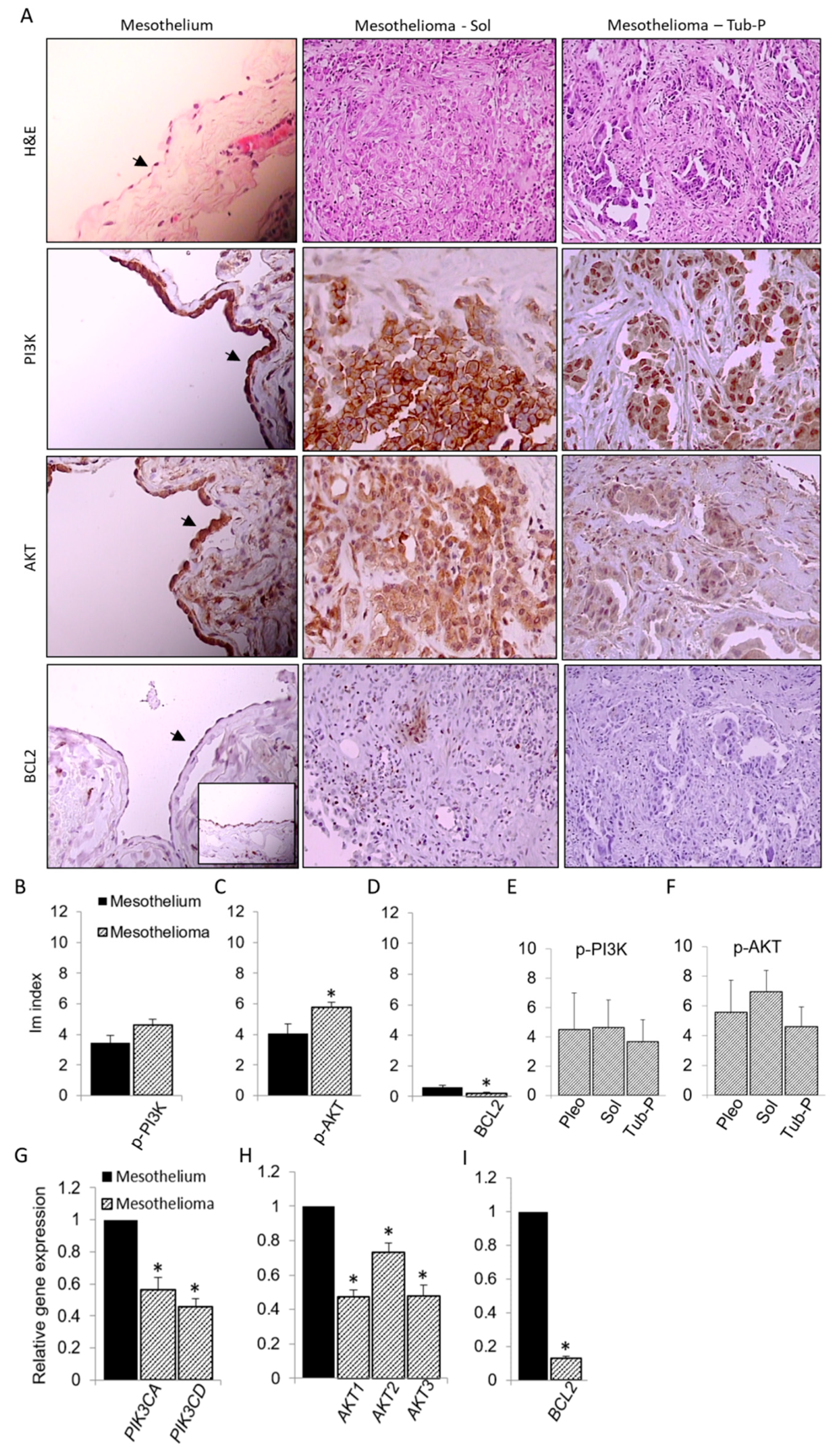
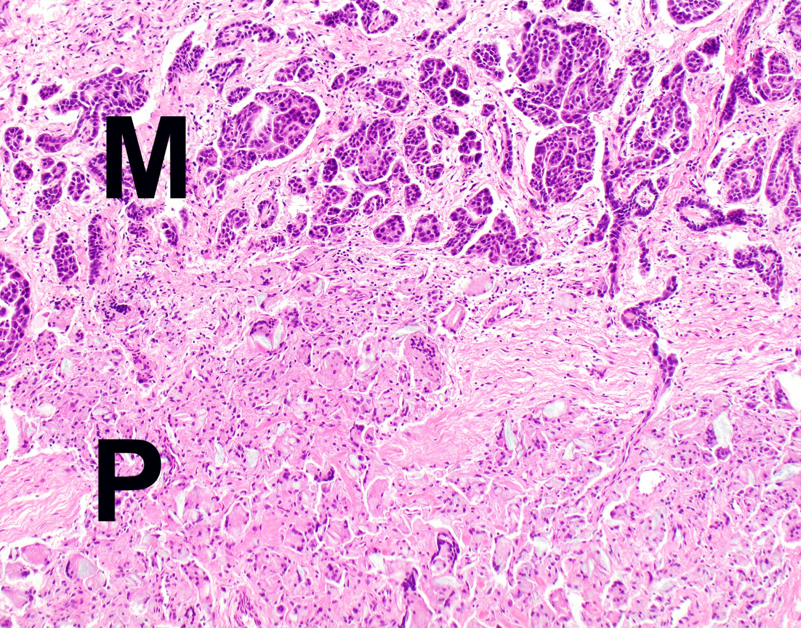
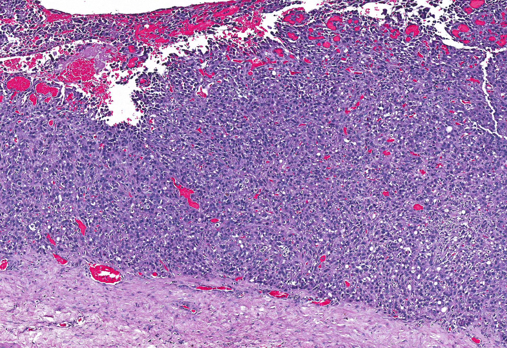
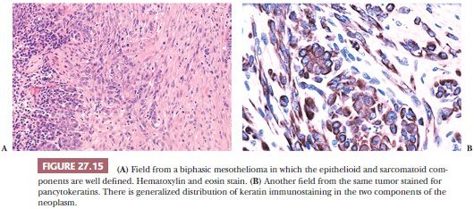
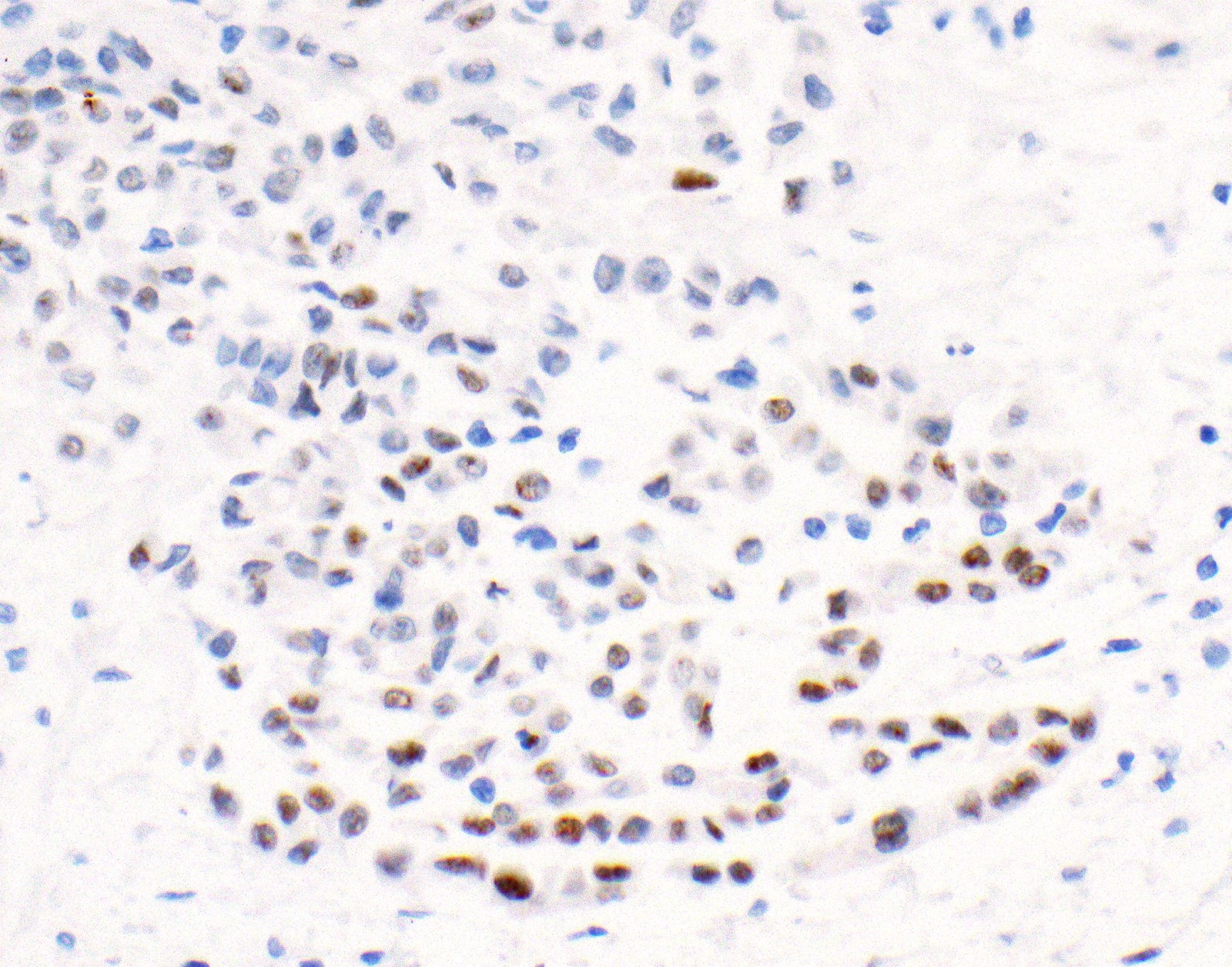
Histopathology falls within the larger field of pathology. Mesothelioma histology involves the study of cancerous mesothelial cells.

Primary Peritoneal Epithelioid Mesothelioma Of Clear Cell Type With A Novel Vhl Gene Mutation A Case Report Sciencedirect
Mesothelioma is a type of cancer that develops from the thin layer of tissue that covers many of the internal organs known as the mesothelium.
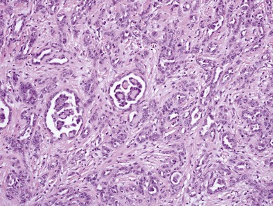
Mesothelioma histology. Observing the anatomy of a particular cancer or how the tumors grow and develop is one way of diagnosing mesothelioma. This form of mesothelioma is comprised of cells which resemble the normal mesothelial cells in that they are arranged in a trabecular fashion. Pathology including histopathology and cytology analyses helps doctors determine the mesothelioma cell type stage and how the cancer is expected to progress.
Mesothelioma histology involves two steps. Mesothelioma pathology is an important aspect of reaching an accurate diagnosis. Histology is a branch of biology that involves the study of cells and tissues.
The most common area affected is the lining of the lungs and chest wall. The tissue samples are sent to a laboratory where an experienced pathologist will review the samples and look for epithelioid sarcomatoid or biphasic mesothelioma cells. This process is part of mesothelioma pathology which involves examining either tissue or fluid to determine if this cancer exists in the body.
The pathology of tumor growth. Mesothelioma pathology provides a full picture of the cancer contributing to a more accurate diagnosis and an informed treatment plan. Well differentiated papillary mesothelioma.
Histologically epithelial mesothelioma cells have polygonal ovoid or cuboidal cell shape. Signs and symptoms of mesothelioma may. The occasional presence of signet ring cells may make it challenging to distinguish this disease from cancers of the lung.
Doctors perform a tissue biopsy on a patient to collect tissue samples. The most obvious anatomical sign of mesothelioma is the. Papillae with myxoid cores each lined by a single mesothelial cell layer invasion is typically not present am j surg pathol 201438990 overall more indolent than peritoneal malignant mesothelioma ann surg oncol 201926852.
Most commonly in the peritoneum rarely pleura and other sites histology. Mesothelioma histology or mesothelioma histopathology is the study of tissue for the presence of mesothelioma. Less commonly the lining of the abdomen and rarely the sac surrounding the heart or the sac surrounding the testis may be affected.
Another subset of this field called histopathology looks specifically at the microstructure of diseased cells and tissues which is what a pathologist will focus on when studying a tissue sample to make a diagnosis. Mesothelioma histology is the study of the function and structure of anatomy including tissues and cells. Histopathology is the study of diseased cells.

Localized Malignant Pleural Mesothelioma Report Of Two Cases Journal Of Thoracic Oncology

Figure 3 From Pathology Of Mesothelioma Semantic Scholar

Malignant Pleural Mesothelioma Clinical Gate
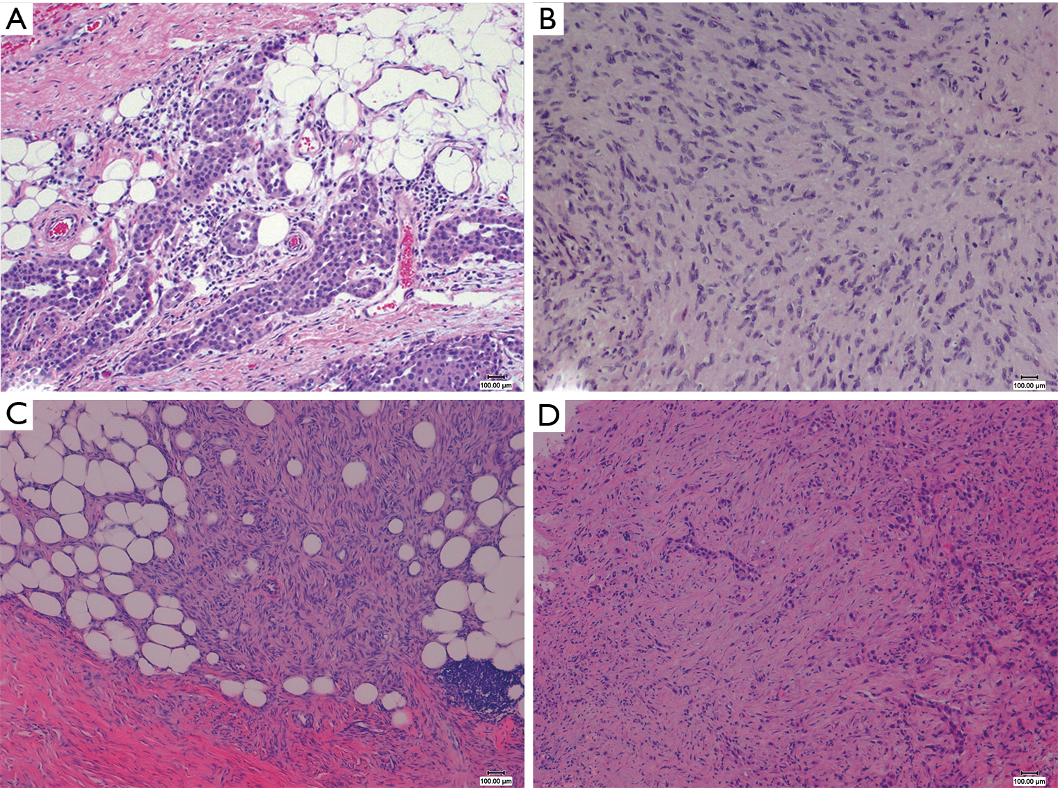
The Pathological And Molecular Diagnosis Of Malignant Pleural Mesothelioma A Literature Review Ali Journal Of Thoracic Disease

Figure 4 From Pathology Of Mesothelioma Semantic Scholar

Challenges And Controversies In The Diagnosis Of Malignant Mesothelioma Part 2 Malignant Mesothelioma Subtypes Pleural Synovial Sarcoma Molecular And Prognostic Aspects Of Mesothelioma Bap1 Aquaporin 1 And Microrna Journal Of Clinical Pathology

