Tag Archives: mesothelioma reactive mesothelial cells

Mesothelioma Reactive Mesothelial Cells

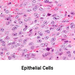
Neoplastic transformation of mesothelial cells results in malignant mesothelioma an aggressive tumor especially the pleura. The morphological evaluation of cytological specimens from body cavity fluids presents difficulties in the differential diagnosis between benign reactive mesothelial rm cells and adenocarcinoma ac or malignant mesothelioma mm.


Mesothelial Images Stock Photos Vectors Shutterstock

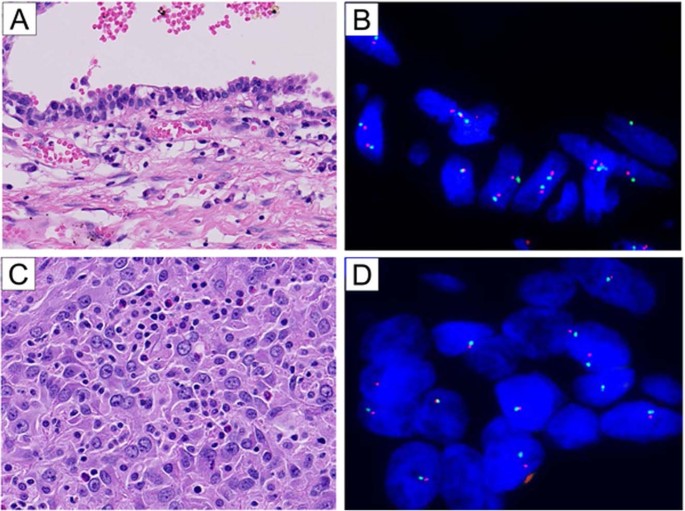
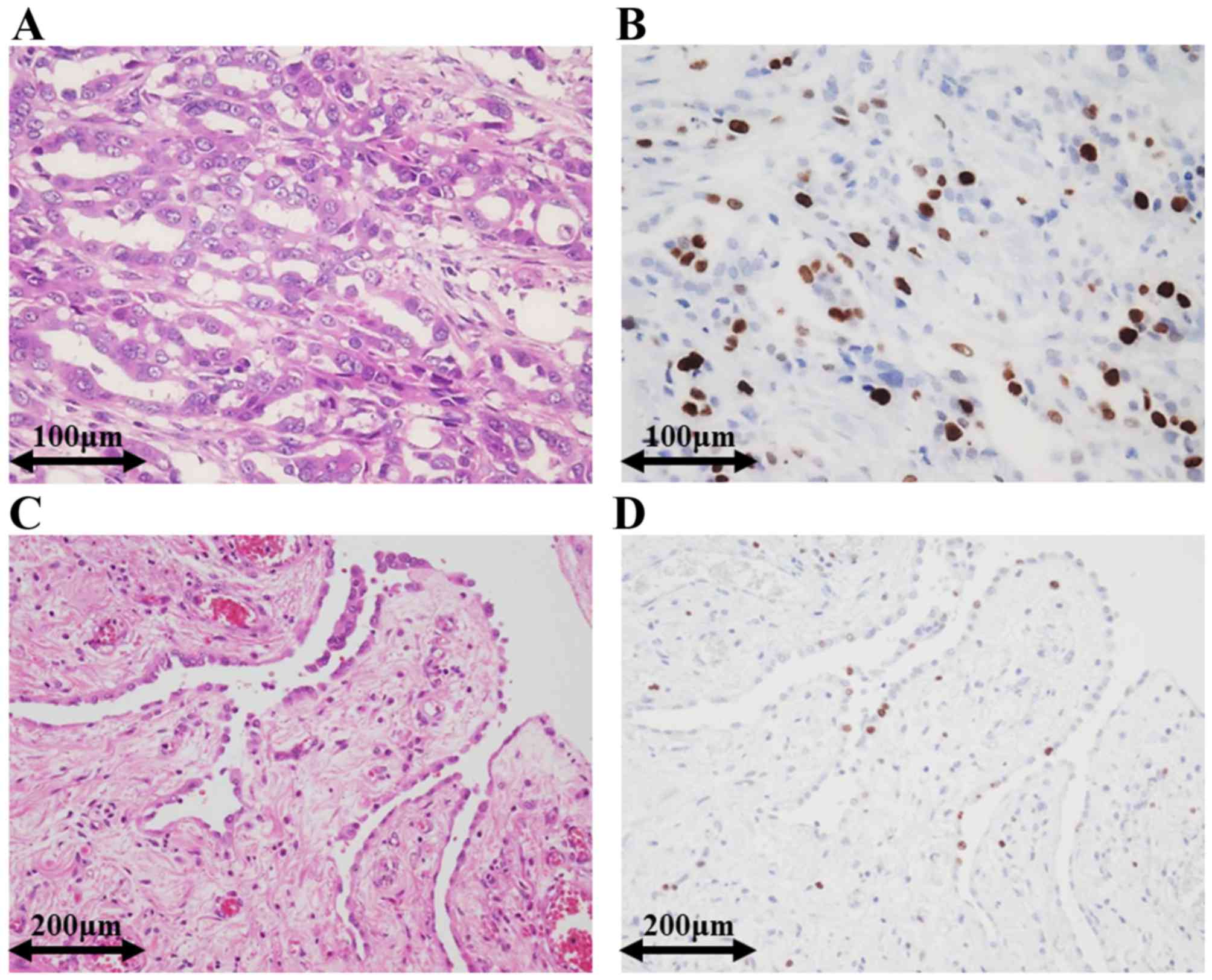
P16 fish followed by immunofluorescence with ema was helpful towards identifying the mesothelioma cells in the cell blocks.

Mesothelioma reactive mesothelial cells. Reactive mesothelial proliferations can occur with infections pneumonia pneumothorax trauma and fluid overload and require a different treatment than malignant mesothelioma. Large nc ratios may be seen in reactive mesothelial cells. Malignant mesothelioma 625 compared with reactive mesothelial proliferation 20 and adenocarcinoma 75.

Immunohistochemical detection of glut 1 can discriminate between reactive mesothelium and malignant mesothelioma. Most adenocarcinoma smears showed a pop. 3 d clusters of cells strongly.

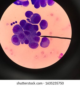
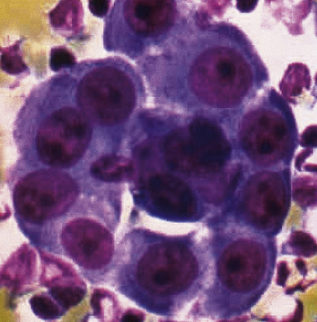
Nc ratio may be normal in mesothelioma. A uniform cell population that is not signicantly different from one another were seen in 100 of cases of malignant mesothelioma as well as in reactive mesothelial proliferation. Reactive mesothelial cells reactive mesothelial cells in pleural fluid reactive mesothelial cells are found when there is infection or inflammation present in a body cavity.

163299 305 26 kato y tsuta k seki k et al. Nuclear membrane irregularies rare. Actually most of the patients with malignant mesothelioma do not get any specific treatment.

Seventeen of the 22 mesothelioma patients 773 showed homozygous deletions of p16 in the tumor tissue and in the atypical mesothelial cells from the cell blocks. Focal macronucleoli are seen in reactive mesothelial cells. The aim of our study was to investigate whether a panel of five dif.

The differential diagnosis of epithelial type mesothelioma from adenocarcinoma and reactive mesothelial proliferation. However if the asbestos exposure is continued over time such heavy volumes of asbestos can overwhelm the mesothelial cells. In the case of mesothelioma when the mesothelial cells are exposed to asbestos fibers the cells ingest these fibers and prevent them from causing harm.

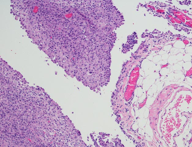
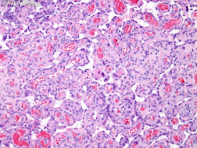
Focal hyperchromasia is seen in reactive mesothelial cells. 1 frank invasion is regarded as the most. The distinction between reactive mesothelial hyperplasia mh and malignant mesothelioma mm may be very difficult based only on histologic and morphologic findings.

Of 217 cases circulated among all members of the uscanadian mesothelioma reference panel there was some disagreement about whether the process was benign or malignant in 22 of cases.


Webpathology Com A Collection Of Surgical Pathology Images


Malignant Mesothelioma In Situ Morphologic Features And Clinical Outcome Modern Pathology


Malignant And Borderline Mesothelial Tumors Of The Pleura Thoracic Key


Https Encrypted Tbn0 Gstatic Com Images Q Tbn 3aand9gctjxi34atmipfik6awy9fwku9hnngvwavizoylai85qzyd 85bh Usqp Cau


Mesothelial Hyperplasia An Overview Sciencedirect Topics


Table 4 From The Use Of Immunohistochemistry To Distinguish Reactive Mesothelial Cells From Malignant Mesothelioma In Cytologic Effusions Semantic Scholar


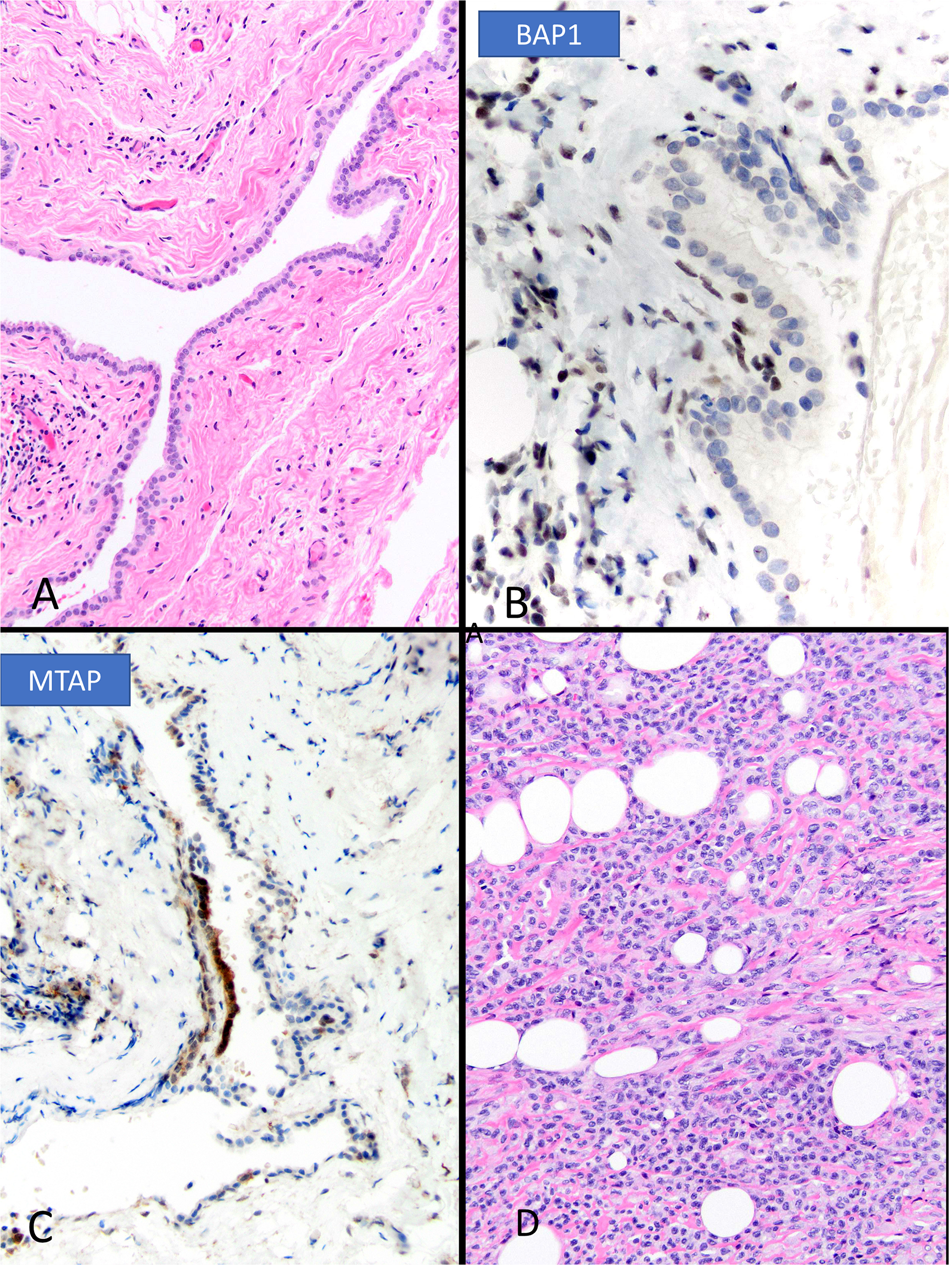
Utility Of Survivin Bap1 And Ki 67 Immunohistochemistry In Distinguishing Epithelioid Mesothelioma From Reactive Mesothelial Hyperplasia

Mesothelioma Cells

Mutations in mesothelial cells caused by asbestos exposure create these variations. Mesothelioma is a type of cancer that develops from the thin layer of tissue that covers many of the internal organs known as the mesothelium.


Hemizygous Loss Of Nf2 Detected By Fluorescence In Situ Hybridization Is Useful For The Diagnosis Of Malignant Pleural Mesothelioma Modern Pathology

Car t cell therapy is an emerging treatment and form of immunotherapy that uses a patients immune system to fight cancers like mesothelioma.

Mesothelioma cells. There are a number of factors that allow a doctor to diagnose the type of mesothelioma that you have and one of them is cell type. Mesothelial cells create a unique layer of paved like cells that line the bodys serious cavities and internal organs. The three major mesothelioma cell types are epithelioid sarcomatoid and biphasic.

Cancer starts when cells start to grow out of control. There are three main types of mesothelioma cells. Mesothelioma cell types explained.

Mesothelioma doctors consider cell type when creating a treatment plan because the cells respond differently to treatment. The most common area affected is the lining of the lungs and chest wall. In the case of mesothelioma when the mesothelial cells are exposed to asbestos fibers the cells ingest these fibers and prevent them from causing harm.

A mesothelioma patients treatment options and prognosis are directly affected by the different types of mesothelioma cells. There are different mesothelioma cells and each of them should be treated with a different treatment. Signs and symptoms of mesothelioma may.

Cells in nearly any part of the body can become cancer. For more information call us today at 888 204 2920. However if the asbestos exposure is continued over time such heavy volumes of asbestos can overwhelm the mesothelial cells.

Doctors will use cell type with mesothelioma type when determining viable treatment options and patient prognosis. The n body contains many different types of cells each of which has a specific function. About 60 percent of all mesothelioma a form of cancer caused by asbestos exposure cases are epithelioid.

The three mesothelioma cell types are epithelioid biphasic and sarcomatoid. The primary function of this layer termed the mesothelium is to provide a slippery non adhesive and protective surface. Malignant mesothelioma is cancer that starts in cells in the linings of certain parts of the body most commonly the linings of the chest or abdomen belly.

Epithelial sarcomatoid and biphasic. Car t cell therapy and mesothelioma. The epithelial mesothelioma cell type makes up more than 50 of all cases.

Researchers genetically modify t cells in a lab and inject them back into the patients body where they prevent or slow the spread of disease. Treatment and prognosis are affected by a patients. Less commonly the lining of the abdomen and rarely the sac surrounding the heart or the sac surrounding the testis may be affected.

Generally the types of mesothelioma cells can be significant in a patients prognosis and cancer treatment plan.


Multiple Sclerosis Drug Effectively Stops Mesothelioma Cells


Detection Of Malignant Mesothelioma Using Nuclear Structure Of Mesothelial Cells In Effusion Cytology Specimens Tosun 2015 Cytometry Part A Wiley Online Library


Ijms Free Full Text Experimental Model Of Human Malignant Mesothelioma In Athymic Mice


Benign Mesothelial Cells With Highly Irregular Nuclear Membranes And Download Scientific Diagram


Malignant Mesothelioma Cells Are Rapidly Sensitized To Trail Induced Apoptosis By Low Dose Anisomycin Via Bim Molecular Cancer Therapeutics


Mesothelioma Cell Types


Can You Identify Each Mesothelioma Cell Type Mesothelioma Guide