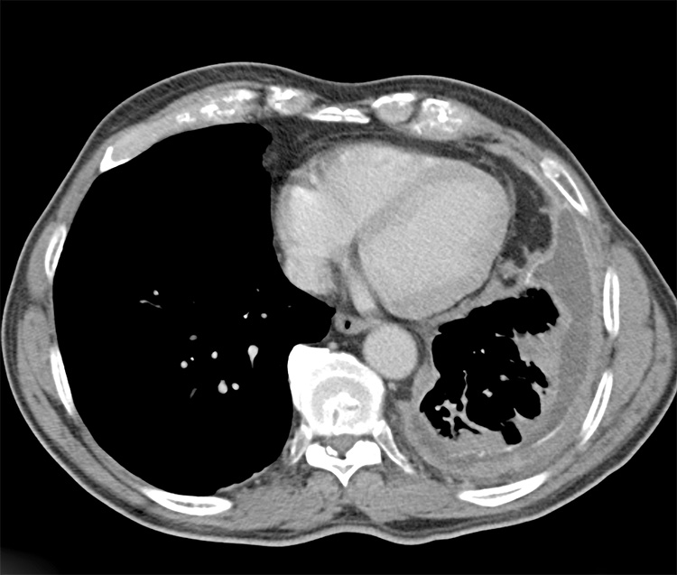
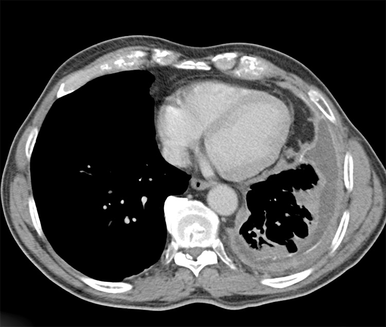
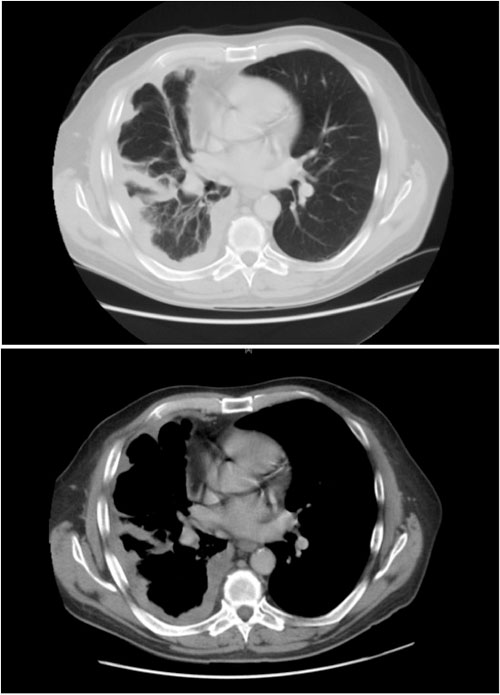
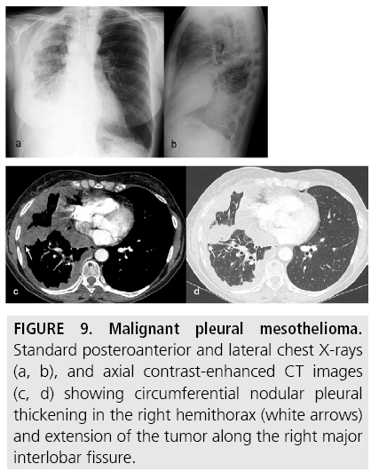
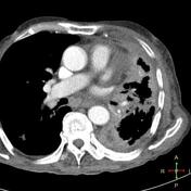
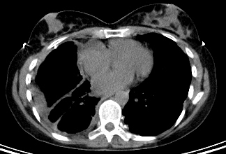
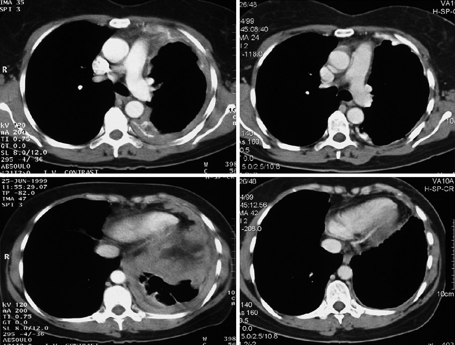
A axial contrast enhanced chest ct scan shows a nodular right sided posterior pleural mass with associated calcification arrow a finding that is consistent with the patients known history of mesothelioma. Information obtained from ct images will.

Computer Tomography Of Patient With Malignant Pleural Mesothelioma Download Scientific Diagram
A ct scan offers a great deal of detail of your chest because it makes a 3d image of your body.

Pleural ct scan mesothelioma. When confronted with the possibility of a deadly disease such as mesothelioma doctors and patients should consider pet scans andor pleural sampling and biopsies in addition to ct scans. Diagnostic ct scan for mesothelioma. Esmo clinical practice guidelines for diagnosis treatment and follow up.
While it is common for ct scans to be taken of just the chests of patients exhibiting mesothelioma symptoms the uk study found that doing so often misses the full extent of the disease. P baas and others on behalf of the esmo guidelines committee. This upper ct scan slice reveals the calcified pleural plaques along the diaphragmatic surface that are associated with asbestos exposure.
Pleural mesothelioma 90 covered in this article. Ultimately a biopsy is the only way to diagnose mesothelioma although the ct scan helps direct when and where to biopsy. In an examination of 249 patients presenting with unilateral pleural effusion a collection of fluid on one side of the chest that is.
Scans can suggest or rule out mesothelioma diagnosis completely while also helping to estimate the extent or stage of mesothelioma. For the accurate diagnosis of pleural mesothelioma and other pleural malignancies a combination of ct scan with positron emission tomography pet is very important. The researchers concluded that relying solely on the ct scan results is not sufficient enough to confirm or negate the presence of pleural malignancy or mesothelioma.
Ascites is seen lateral to the liver. It takes pictures from different angles. When a person goes to the doctor with early signs of malignant pleural mesothelioma a ct scan of the chest is often the next step in making a diagnosis.
Furthermore although less effective for detecting peritoneal mesothelioma ct scans are still the most useful imaging study for diagnosing malignant pleural mesothelioma. The imaging process begins when you lie on a table over which the device is attached. Mesothelioma also known as malignant mesothelioma is an aggressive malignant tumor of the mesothelium.
More is better a new report out of the uk suggests that a thorough diagnostic ct scan for pleural mesothelioma should include images of the abdomen and pelvis as well as the chest. Computed tomography ct scan in a male veterans administration patient with a history of asbestos exposure and an enlarging abdominal girth. This allows doctors to see behind any fluid buildups that may block vital visual signs of asbestos cancer such as pleural thickening or tumors.
Most tumors arise from the pleura and so this article will focus on pleural mesothelioma. A ct scan is a test that uses x rays and a computer to create detailed pictures of the inside of your body. Given the presence of the mesothelium in different parts of the body mesothelioma can arise in various locations 17.
Imaging abdomen and pelvis essential in suspected pleural mesothelioma.
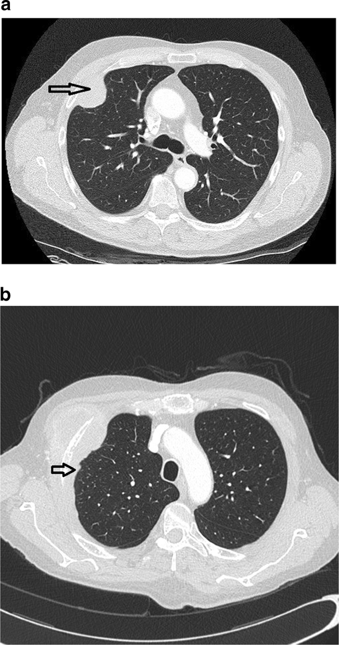
Localized Malignant Mesothelioma An Unusual And Poorly Characterized Neoplasm Of Serosal Origin Best Current Evidence From The Literature And The International Mesothelioma Panel Modern Pathology

Localized Malignant Pleural Sarcomatoid Mesothelioma Misdiagnosed As Benign Localized Fibrous Tumor Kim Journal Of Thoracic Disease
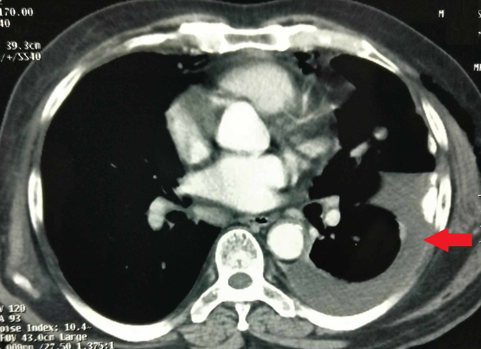
Cureus Prostate Carcinoma And Pleural Mesothelioma An Extremely Rare Co Occurrence

Malignant Pleural Effusion Pulmonology Advisor

Radiological Review Of Pleural Tumors Sureka B Thukral Bb Mittal Mk Mittal A Sinha M Indian J Radiol Imaging

Figure I From Malignant Pleural Mesothelioma Semantic Scholar

Pleura Chest Wall And Diaphragm Chest Radiology The Essentials 2nd Edition
