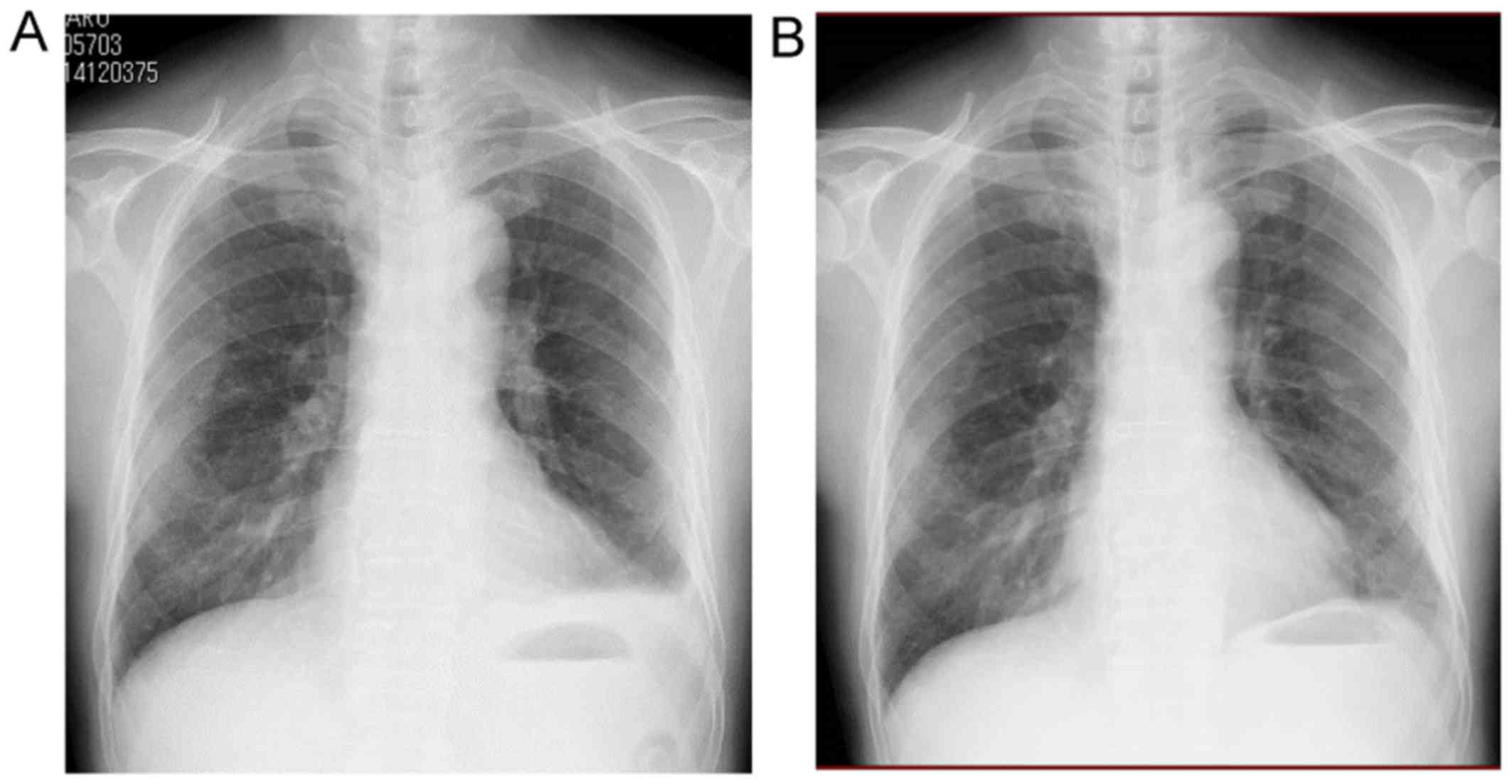
Tag Archives: pleural mesothelioma cxr

Mesothelioma Cxr

Before the x ray the technologist may ask you to remove your clothing from the waist up. It can also show up a rim of solid tumour around the lung.


Decortication

Less commonly the lining of the abdomen and rarely the sac surrounding the heart or the sac surrounding the testis may be affected.
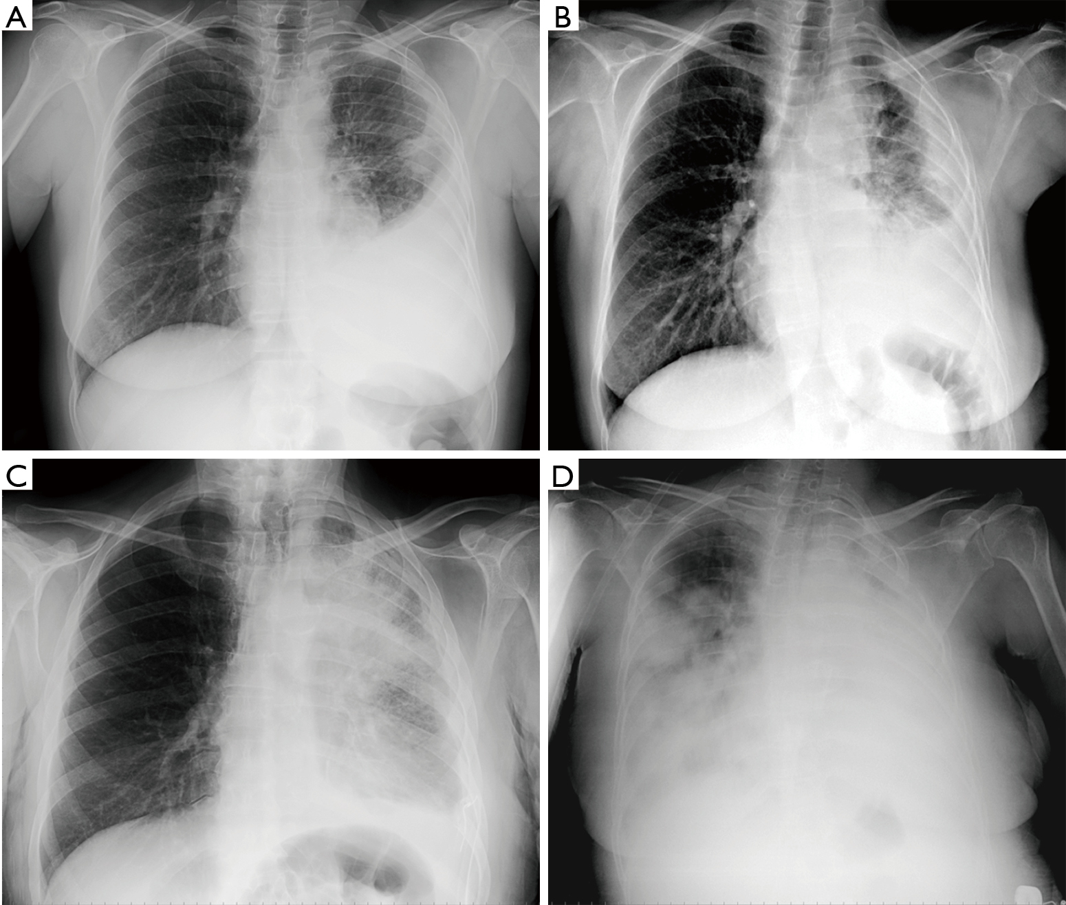
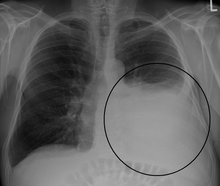
Mesothelioma cxr. Ct also gives important information regarding invasion of the chest wall and surrounding structures. Primary tumor cannot be assessed. The most common area affected is the lining of the lungs and chest wall.

However a chest x ray has limited usefulness because the images findings are nonspecific. Usually you also need an ultrasound of your tummy. But computed tomography ct is the imaging technique of choice for charactering pleural masses.

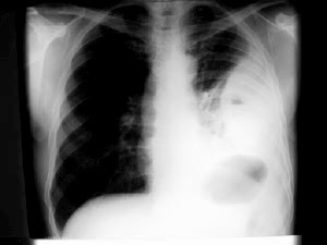
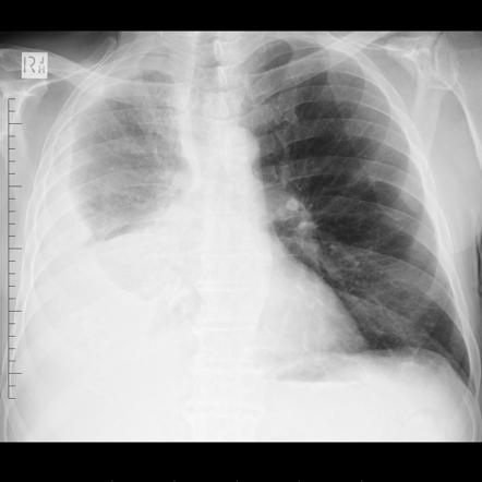
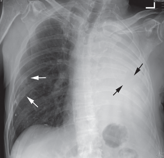
Pleural mesothelioma 90 covered in this article. Chest radiograph cxr of a 50 year old male who presented with chronic dry cough of a few months duration. Mediastinal and diaphragmatic pleura visceral pleura.

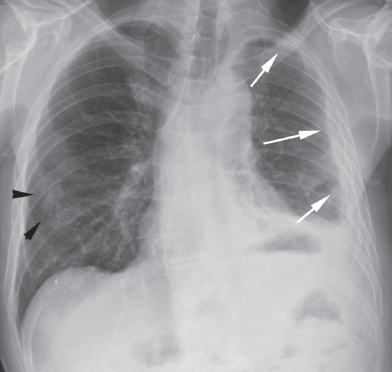
Signs and symptoms of mesothelioma may. The radiograph shows lobulated pleural based masses in the right hemithorax peripherally white arrows. A chest x ray is performed by a radiology technologist.

Mesothelioma is a rare type of cancer. Pleural biopsy confirmed malignant mesothelioma. A chest x ray for mesothelioma patients will inevitably show irregularities.

Mesothelioma is a type of cancer that develops from the thin layer of tissue that covers many of the internal organs known as the mesothelium. Most tumors arise from the pleura and so this article will focus on pleural mesothelioma. No evidence of primary tumor.

An x ray of your tummy abdomen might show up a swelling or fluid collecting in the tummy. Mesothelioma also known as malignant mesothelioma is an aggressive malignant tumor of the mesothelium. Given the presence of the mesothelium in different parts of the body mesothelioma can arise in various locations 17.

The chest x ray cannot delve deep into the tissues and reveal irregularities that are picked up by more advanced imaging platforms. One of the usual first steps to a mesothelioma diagnosis is a chest x ray. A chest x ray can show up fluid collecting in the lining of your lung the pleura.

A chest x ray may show a buildup of fluid in the lining around the lung and is a quick procedure that requires no preparation. Chest x ray is the initial screening test for the mesothelioma like all other the chest diseases. It typically develops in the thin membrane that separates the lung from the chest wall called the pleurait can also arise less commonly along the abdominal cavity inner or peritoneal liningmesothelioma can result in breathing difficulties chest pain and fever.

Involving ipsilateral parietal pleura inc. Below is the eighth edition of the tnm staging system for malignant pleural mesothelioma which was published in 2018 1. Involving any of the ipsilateral pleural surfaces with at least one of the.

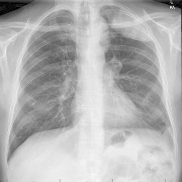
No pleural effusion is present.


Clinical Diagnosis Of Malignant Pleural Mesothelioma Bianco Journal Of Thoracic Disease


Malignant Pleural Mesothelioma With Pleural Plaques A Case Report


Medpix Case Malignant Mesothelioma


Mesothelioma Radiology Case Radiopaedia Org


Malignant Mesothelioma Harrisons Manual Of Oncology 2nd Ed
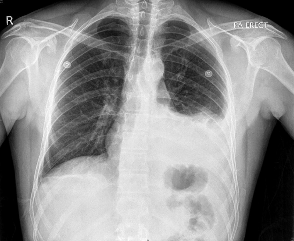


Malignant Mesothelioma Clinical And Imaging Findings Springerlink
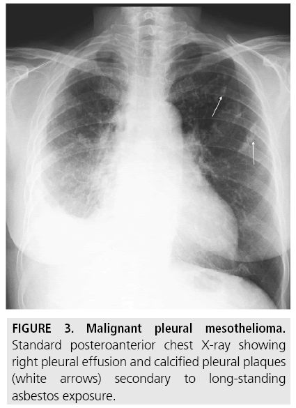


Diagnostic Imaging And Workup Of Malignant Pleural Mesothelioma
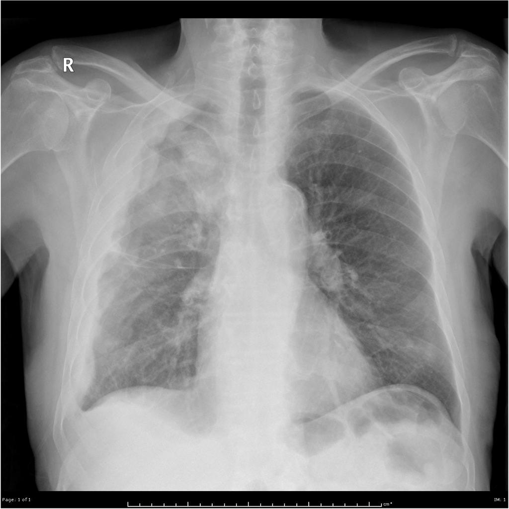
Pleural Mesothelioma Cxr

Given the presence of the mesothelium in different parts of the body mesothelioma can arise in various locations 17. Mesothelioma is a malignant neoplasm originating from pleural or peritoneal surfaces.


Chest X Ray Open I

Involving any of the ipsilateral pleural surfaces with at least one of the.

Pleural mesothelioma cxr. Most tumours arise from the pleura and so this article will focus on pleural mesothelioma. Involving any of the ipsilateral pleural surfaces with at least one of the. Pleural plaques are strongly associated with inhalational exposure to asbestosthere is an extremely long latency typically 20 to 30 years after the onset of exposure 1as asbestos is primarily used in construction and machinery environments asbestos related diseases in general including pleural plaques are primarily seen in men.

Mediastinal and diaphragmatic pleura visceral pleura. Primary tumor cannot be assessed. Here we present cxr from 64 year old hispanic male who was diagnosed with advanced mesothelioma.

Below is the eighth edition of the tnm staging system for malignant pleural mesothelioma which was published in 2018 1. Involving ipsilateral parietal pleura inc. This article summarises the epidemiology and pathogenesis of malignant pleural mesothelioma before describing some key factors.

Mediastinal and diaphragmatic pleura visceral pleura. The most common mesothelioma finding on radiographs is unilateral concentric plaque like or nodular pleural thickening. Mesothelioma is a type of cancer that develops from the thin layer of tissue that covers many of the internal organs known as the mesothelium.

Mesothelioma also known as malignant mesothelioma is an aggressive malignant tumor of the mesothelium. Malignant pleural mesothelioma is an aggressive malignancy of the pleural surface predominantly caused by prior asbestos exposure. A common result of asbestos exposure.

Below is the eighth edition of the tnm staging system for malignant pleural mesothelioma which was published in 2018 1. Pleural plaques are areas of thickened tissue in the pleura around the lungs. Less commonly the lining of the abdomen and rarely the sac surrounding the heart or the sac surrounding the testis may be affected.

Involving ipsilateral parietal pleura inc. Signs and symptoms of mesothelioma may. No evidence of primary tumour.

There is a global epidemic of malignant pleural mesothelioma underway and incidence rates are predicted to peak in the next few years. No evidence of primary tumor. They are the most common sign of asbestos damage to the chest cavity but may not develop for decades after exposure.

Primary tumour cannot be assessed. Pleural mesothelioma 90 covered in this article. Mesothelioma also known as malignant mesothelioma is an aggressive malignant tumour of the mesothelium.

Pleural effusions are common and may obscure the presence of the underlying pleural thickening. Given the presence of the mesothelium in different parts of the body mesothelioma can arise in various locations 17. Plaques do not typically cause symptoms and may or may not develop into malignant pleural mesothelioma.

The most common area affected is the lining of the lungs and chest wall. This condition is usually associated with occupational exposure to asbestoswagner et al connected asbestos to mesothelioma in a classic 1960 study of 33 patients with mesothelioma who were exposed to asbestos in a mining area in south africas north western cape province. Most tumors arise from the pleura and so this article will focus on pleural mesothelioma.

Pleural mesothelioma 90 covered in this article.


Malignant Pleural Mesothelioma With Pleural Plaques A Case Report


Mesothelioma Radiology Case Radiopaedia Org


Early Detection Of Malignant Pleural Mesothelioma Mahmoud Hf Egypt J Bronchol


Medpix Case Malignant Mesothelioma


Double Cancer Comprising Malignant Pleural Mesothelioma And Squamous Cell Carcinoma Of The Lung Treated With Radiotherapy A Case Report


Pleural Effusion Wikipedia


Plain Chest X Ray Findings In The Malignant Pleural Mesothelioma Download Table