Tag Archives: pleural thickening mesothelioma x ray

Pleural Mesothelioma X Ray

They are a good way to look at bones and can show changes caused by cancer or other medical conditions. Ct scans for example arent very effective in detecting peritoneal mesothelioma because gas in the intestines can be mistaken for tumors.


Mmcts Extended Pleurectomy Decortication For The Treatment Of Malignant Pleural Mesothelioma

X ray images may be helpful for detecting pleural effusions excess fluid around the lungs.

Pleural mesothelioma x ray. Doctors may choose to perform a chest x ray ct scan mri or another diagnostic test to look for cancer. Imaging tests are used to look for tell tale signs of pleural mesothelioma such as thickening of the pleural lining pleural effusions and tumors growing around the lung area. People diagnosed with mesothelioma have aggressive cancer that is caused by asbestos exposure.

Pleural mesothelioma is a cancer of the pleural cavity which is a thin membrane between the chest wall and lung cavity. However the presence of a pleural effusion could also indicate other abnormalities in the lung or the pleura. However some tests arent as effective as others for different types of this cancer.

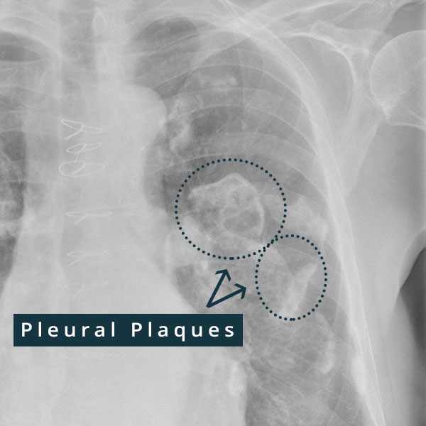
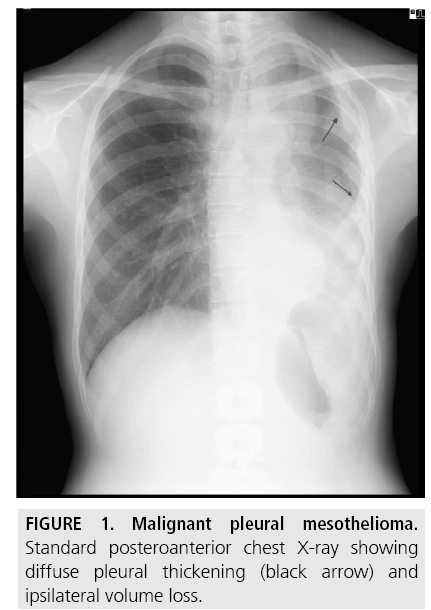
The x ray may reveal pleural thickening commonly seen after asbestos exposure and increases suspicion of mesothelioma. Most tumors arise from the pleura and so this article will focus on pleural mesothelioma. Diagnosing pleural mesothelioma often consists of multiple tests.

An x ray is a test that uses small doses of radiation to take pictures of the inside of your body. You will learn can a chest x ray show mesothelioma and much more. Pleural mesothelioma 90 covered in this article.

Given the presence of the mesothelium in different parts of the body mesothelioma can arise in various locations 17. Esmo clinical practice guidelines for diagnosis treatment and follow up. One or more imaging scans such as an x ray or ct scan may be performed first to identify tumors or metastasis spreading of diseaseif a tumor is detected blood tests may be performed to look for certain biomarkers high levels of specific substances in the blood which can help differentiate mesothelioma from other conditions.

If a large amount of fluid is present abnormal cells may be detected by cytopathology if this fluid is aspirated with a syringe. A ct or cat scan or an mri is usually performed. Although a chest x ray does not provide enough information to confirm a diagnosis it can reveal early signs of mesothelioma such as fluid in the lungs.

A computerized tomography ct scan can create a 3d image that allows tumors to be easily detected. X rays and ct scans are the most common imaging tests used for mesothelioma. The first step in diagnosing mesothelioma is imaging tests such as an x ray or a computed tomography ct scan.

Mesothelioma also known as malignant mesothelioma is an aggressive malignant tumor of the mesothelium. A chest x ray is commonly the first step to achieving a diagnosis of pleural mesothelioma. If the test results determine the possible presence of cancerous tumors doctors likely will perform a biopsy.

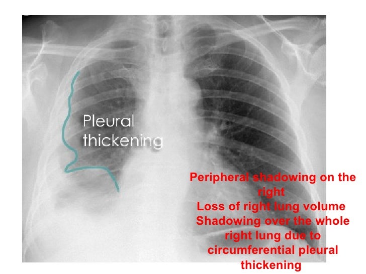
One of the initial signs of mesothelioma is a thickening of the lung or pleural thickening that can be seen on a chest x ray. This cancer is incurable but patients who are diagnosed early have a much greater life expectancy. In many cases chest x rays are helpful for mesothelioma surveillance but even in ideal circumstances can only provide limited information.

P baas and others on behalf of.


Https Encrypted Tbn0 Gstatic Com Images Q Tbn 3aand9gcr0g0ntodbc44xvqx5g6vbit5cjyk9qwdstb58hb1e Usqp Cau


Medpix Case Malignant Mesothelioma


Pleural And Peritoneal Mesothelioma Imaging Findings On Ct And Fdg Pet Ct Sciencedirect
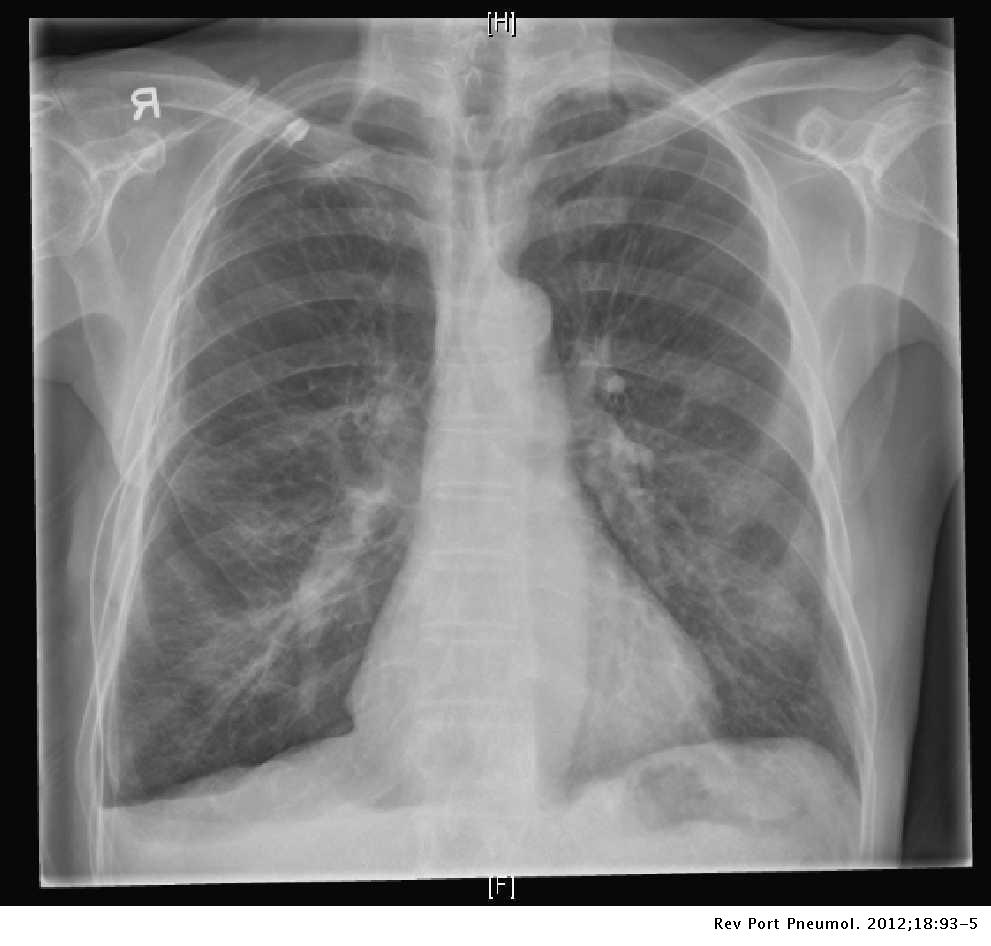


Malignant Pleural Mesothelioma Presenting With A Spontaneous Hydropneumothorax A Report Of 2 Cases Pulmonology


Https Encrypted Tbn0 Gstatic Com Images Q Tbn 3aand9gctu5wwfkxxwsgbbkfauk0qb8mgvh Qb4amhb6eivjpuelgmzrfs Usqp Cau


Biphasic Mesothelioma Chest X Ray Film A Showing A Right Basal And Download Scientific Diagram
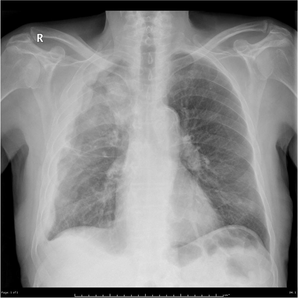


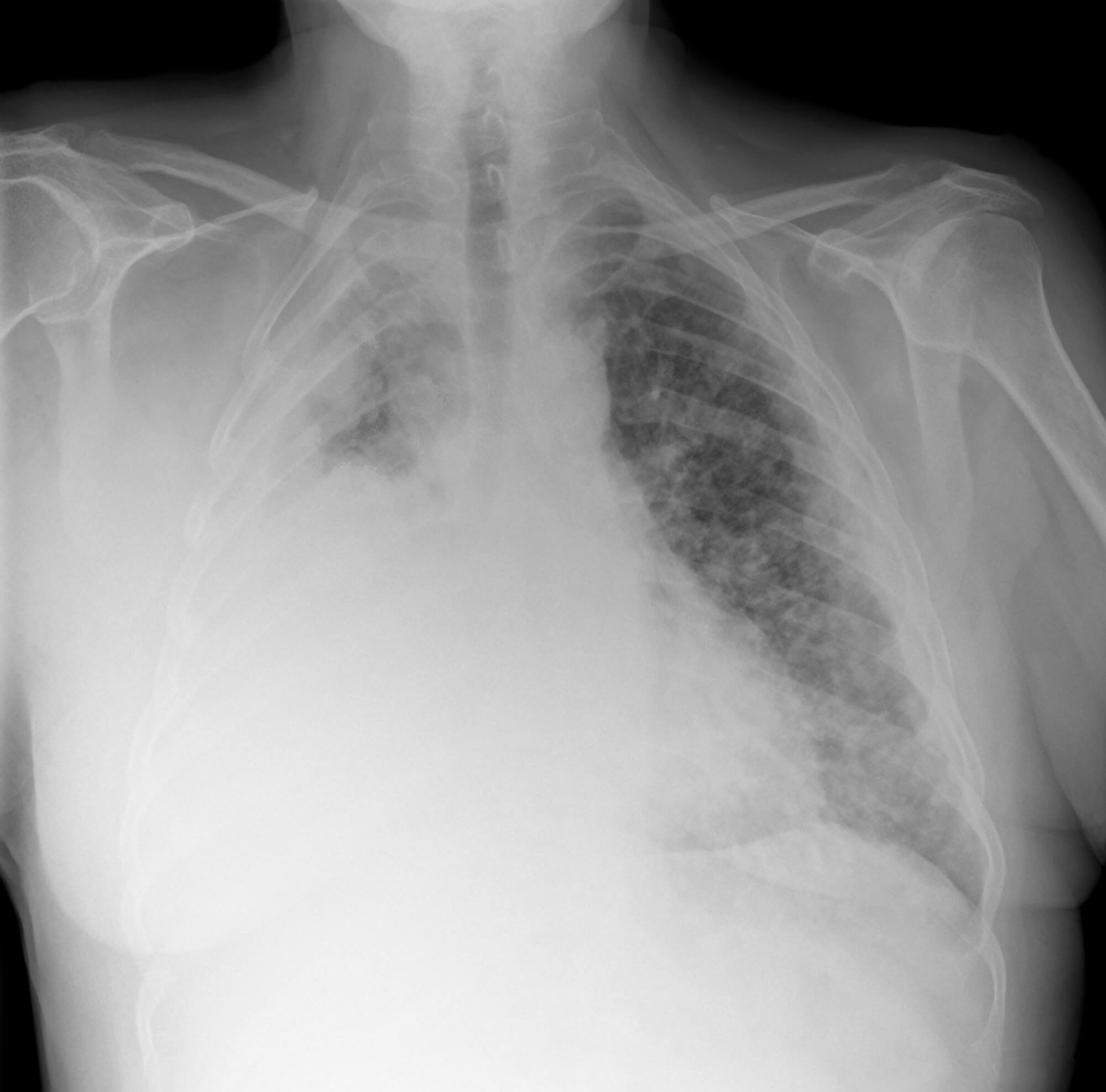
Benign Pleural Thickening Radiology Key

Mesothelioma Pleural Thickening

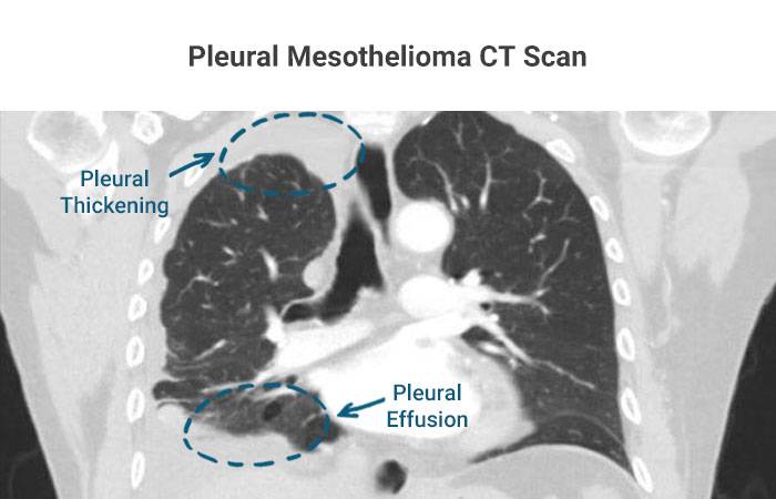
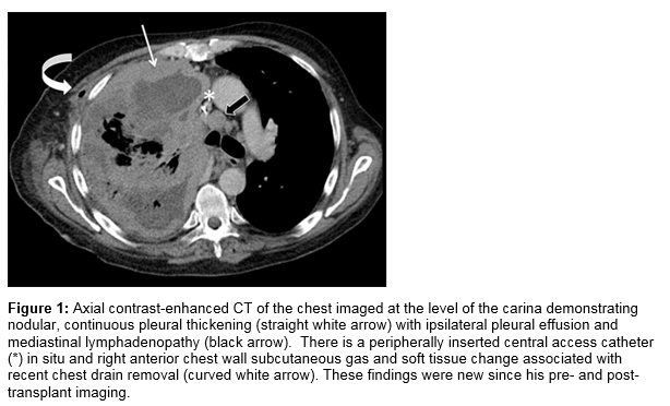
Pleural mesothelioma with effusion and thickening in the right lung. Our expert asbestos disease solicitors have helped hundreds of clients to secure the maximum possible compensation for their pleural thickening claims.


Mr Harrison S Story Pleural Thickening Case Thompsons Solicitors

Treatment tissue biopsy or computerized tomography ct scans as well as tests of a patients pulmonary function may be used in diagnosing suspected pleural thickening.

Mesothelioma pleural thickening. If you are experiencing pleural thickening but have not yet been diagnosed with mesothelioma you may yet have a while to live perhaps years to decades depending on your condition. Pleural thickening may be a symptom of mesothelioma and related conditions or it may be benign. Pleural thickening and mesothelioma.

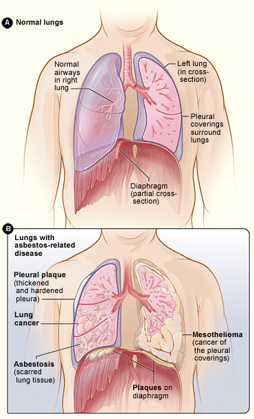
This scarring also known as fibrosis restricts lung function and may cause chest pain and difficulty breathing. Pleural thickening is one of the symptoms of pleural mesothelioma by far the most common form of the diseasethe tissue surrounding the lungs andor lining the chest wall develops inflammation thickens as abnormal cells are generated by the presence of asbestos fibers. However it can be a sign of asbestos exposure.

Because catching mesothelioma in an early stage can lead to a wider range of treatment options patients with asbestos related pleural thickening should seek regular cancer screening. Physicians may be able to obtain an early diagnosis for mesothelioma patients with pet and ct scans. Pleural thickening in the patient does not confirm a mesothelioma diagnosis.

Diffuse pleural thickening is a disease that causes the pleura layers of tissue around the lungs to thicken. Benign pleural thickening is not caused by cancer and will typically not impede the normal functioning of the lungs. Pleural thickening is not deadly on its own but rather is usually a bad sign because it is a precursor to mesothelioma.

There are a number of conditions that may indicate a persons history of asbestos exposure and the potential of a mesothelioma diagnosis including pleural plaques pleural effusions and pleural thickening. In some cases pleural thickening can be benign. It causes difficulty breathing shortness of breath and chest pains.

The presence of pleural thickening is not enough to confirm a pleural mesothelioma diagnosis but it can be a sign of serious and significant asbestos exposure. Pleural thickening is a condition triggered by asbestos exposure that causes the pleural lining of the lungs known as the pleura to thicken with scar tissue. Patients diagnosed with pleural thickening also have an increased risk of developing other asbestos related diseases such as asbestos related lung cancer and the cancer mesothelioma.

For instance pleural thickening may be a sign of malignant mesothelioma cancer. Pleural thickening is also present in metastatic adenocarcinoma as well as mesothelioma a rare cancer of the pleura that is related to asbestos exposure. Pleural thickening is also often an indicator of a serious underlying condition.


Asbestos Related Diseases Wikipedia


Figure 1 From Malignant Pleural Mesothelioma Role Of Ct Mri And Pet Ct In Staging Evaluation And Treatment Considerations Semantic Scholar


Thorax 8 2 Pleural Space Case 8 2 2 Benign And Malignant Pleural Thickening Ultrasound Cases


Obliteration Of Left Costophrenic Angle With Opaque Shadow Seen Along The Chest Wall Suggestive Of Lamellar Pleural Eff Mesothelioma Pleural Effusion Treatment


Malignant Mesothelioma Iii Ct


Asbestos Related Pleural Disease Pulmonary Disorders Merck Manuals Professional Edition


Assessment Of 18f Fluorodeoxyglucose Dual Head Gamma Camera In Asbestos Lung Diseases European Respiratory Society

Mesothelioma X Ray

X rays and ct scans are the most common imaging tests used for mesothelioma. One of the initial signs of mesothelioma is a thickening of the lung or pleural thickening that can be seen on a chest x ray.


Malignant Mesothelioma Imaging Overview Radiography Computed Tomography

An x ray provides a flat 2d image of bones and soft tissue.

Mesothelioma x ray. Radiation therapy is a severe dose of x rays which are directed to the cancerous cells. If you cant stand you can have it sitting or lying on the x ray couch. People diagnosed with mesothelioma have aggressive cancer that is caused by asbestos exposure.

You may first have a chest x ray. A chest ct is a 3 dimensional x ray that shows lung and pleural abnormalities in detail. Radiation therapy also referred to as x ray therapy is used to target and destroy individual tumor cells.

There are five primary types of imaging tests used during a mesothelioma diagnosis including x rays mri scans ct scans pet scans and ultrasounds. The importance of imaging. You usually have a chest x ray standing up against the x ray machine.

However some tests arent as effective as others for different types of this cancer. Often your doctor will order a computed tomography ct scan of your chest as well. This cancer is incurable but patients who are diagnosed early have a much greater life expectancy.

Mesothelioma x ray and imaging scans help specialists find malignant tumors and make more accurate diagnosesimaging also helps guide biopsies and treatment and track the progress of the disease. Given the presence of the mesothelium in different parts of the body mesothelioma can arise in various locations 17. If a large amount of fluid is present abnormal cells may be detected by cytopathology if this fluid is aspirated with a syringe.

Ct scans for example arent very effective in detecting peritoneal mesothelioma because gas in the intestines can be mistaken for tumors. For x rays of other areas of the body the best position is usually lying down on the x ray couch. A chest x ray can help guide physicians towards a mesothelioma diagnosis but cannot definitively diagnose a patient.

They can be helpful for detecting any abnormalities especially for those at high risk for developing mesothelioma such as patients that have smoked or had significant asbestos exposure in the past. Imaging is important in diagnosing mesotheliomait will provide information such as the extent of disease in the original organ and also show if the cancer has spread to. You will learn can a chest x ray show mesothelioma and much more.

Mesothelioma also known as malignant mesothelioma is an aggressive malignant tumor of the mesothelium. A high dose of x rays will be administered as a mesothelioma treatment option in a variety of ways. As the cancerous cells die the main tumor shrinks.

A ct or cat scan or an mri is usually performed. Most tumors arise from the pleura and so this article will focus on pleural mesothelioma. Pleural mesothelioma 90 covered in this article.

The radiographer lines the machine up to make sure its in the right place. Imaging scans are non invasive and act as a stepping stone in the diagnostic process for mesothelioma patients. In general individuals at risk for and with symptoms of mesothelioma are referred to a specialist in lung diseases called a pulmonologist.

The x ray may reveal pleural thickening commonly seen after asbestos exposure and increases suspicion of mesothelioma.


Asbestos When The Dust Settles An Imaging Review Of Asbestos Related Disease Radiographics


Postoperative X Ray Of The Chest After Rp And Thoracic Shaping Download Scientific Diagram
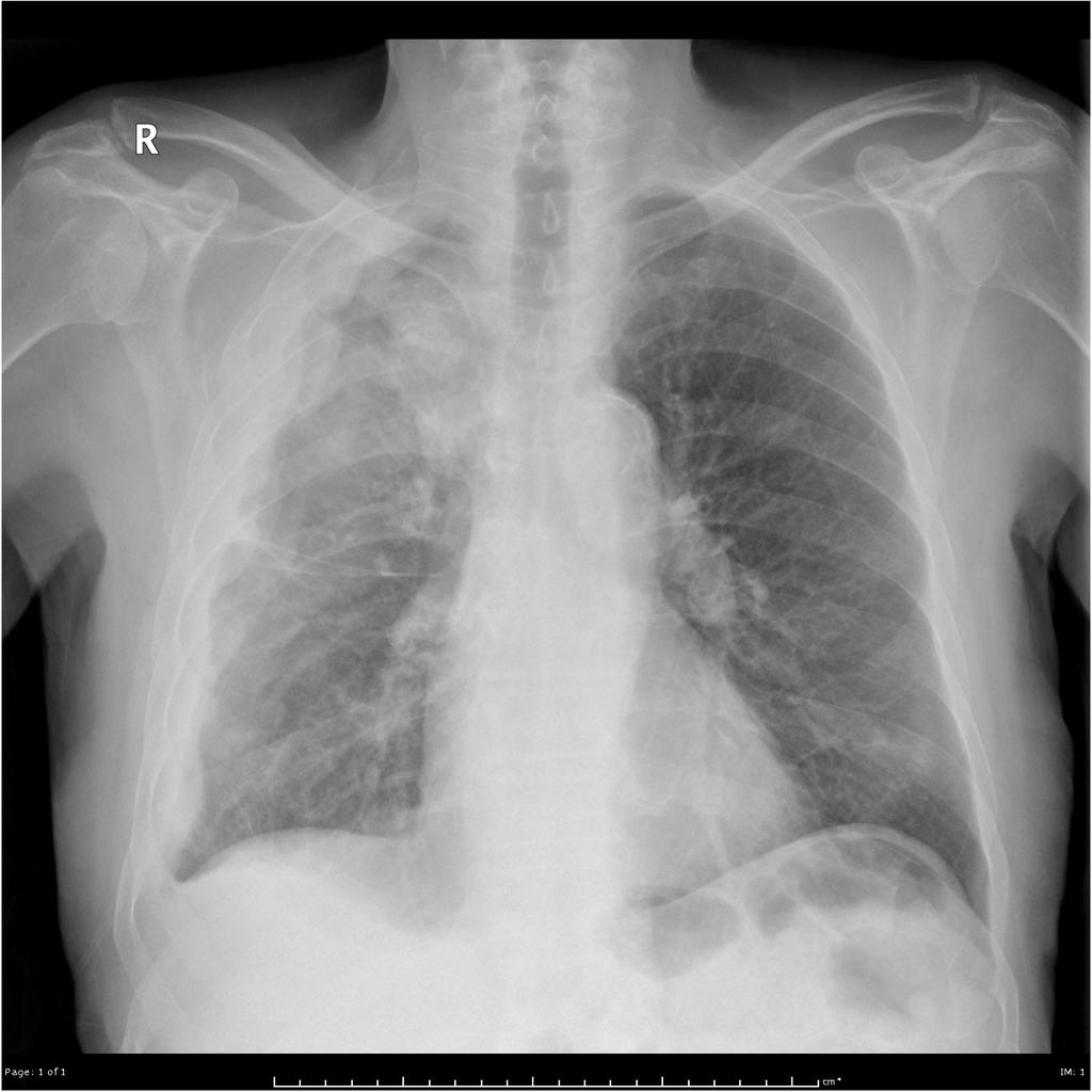


View Of Malignant Mesothelioma The Southwest Respiratory And Critical Care Chronicles


Malignant Pleural Mesothelioma Presenting With A Spontaneous Hydropneumothorax A Report Of 2 Cases Pulmonology


A Shows The Chest X Ray Of A Mesothelioma Patient With A Trapped Lung Download Scientific Diagram


Mesothelioma Lung Cancer X Ray Iphone X Case For Sale By Science Photo Library


Pleural Plaques Causes Symptoms Diagnosis Treatment

Pleural Thickening Mesothelioma X Ray

If a patient is supine then a pleural effusion layers along the posterior aspect of the chest cavity and becomes difficult to see on a chest x ray. If the patient is upright when the x ray is taken then fluid will surround the lung base forming a meniscus a concave line obscuring the costophrenic angle and part or all of the hemidiaphragm.


Cxr Pneumothorax Pleural Thickening

Some clinical studies show that when compared to an x ray a ct scan is better able to see pleural thickening asbestosis and pleural plaques.

Pleural thickening mesothelioma x ray. The condition is most often diagnosed with a chest x ray but a diagnosis from a ct scan is better. A physician will check for physical symptoms such as altered breathing sounds. Given the presence of the mesothelium in different parts of the body mesothelioma can arise in various locations 17.

Often pleural thickening is diagnosed alongside its cause. Although it may not show everything necessary for diagnosis. A ct scan which is also used to diagnose asbestosis and pleural plaques can confirm the condition earlier.

Another finding is diffuse pleural thickening in the absence of pleural effusion. Healthy pleurae are not visible on an x ray but thickening at the very edges of the lungpleura may be. These scans are high resolution and allow doctors to distinguish between pleural thickening and pleural plaques.

This cancer is incurable but patients who are diagnosed early have a much greater life expectancy. An x ray can reveal thickening of the pleura but a ct scan is more likely to show thickening in better detail. Asbestos exposure can cause diffuse pleural thickening which results from lesions forming on the lung lining the pleura.

Methods used to detect and diagnose pleural thickening include. One of the initial signs of mesothelioma is a thickening of the lung or pleural thickening that can be seen on a chest x ray. People diagnosed with mesothelioma have aggressive cancer that is caused by asbestos exposure.

The most common way to diagnose pleural thickening is with x rays mris or ct scans. Ct scans also can see earlier signs of pleural thickening with scar tissue just 1 mm or 2 mm thick. Pleural thickening can be a sign of serious conditions such as malignant mesothelioma and lung cancer or it can be benign.

Pleural mesothelioma 90 covered in this article. Pleural thickening as well as pleural effusion and pleural classification as well as lymphonodi around hillus or mediastinum look more clearly and can be. You will learn can a chest x ray show mesothelioma and much more.

In terms of mesothelioma x ray findings may show a pleural mass on one side of the lung with pleural effusion or fluid. Pet and mri scans provide even further detail and may be used to differentiate pleural thickening from malignant mesothelioma. Mesothelioma also known as malignant mesothelioma is an aggressive malignant tumor of the mesothelium.

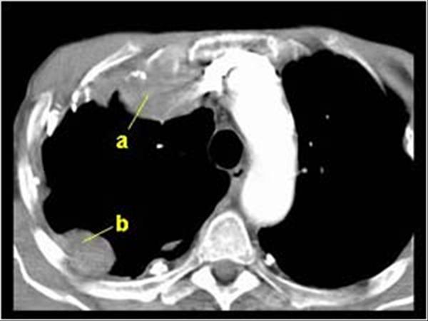
A ct or computed tomography scan gives a clearer picture of what is happening in the lungs and pleura. Most tumors arise from the pleura and so this article will focus on pleural mesothelioma. The pleural thickening in mesothelioma usually is in the part of lung basal or medio basal hemi 75 thorax3678910 chest ct scan in the mesothelioma shows more clearly than chest x ray.

A chest x ray is a typical first screening.


Pleural Tuberculosis A Key Differential Diagnosis For Pleural Thickening Even Without Obvious Risk Factors For Tuberculosis In A Low Incidence Setting Bmj Case Reports


Non Malignant Asbestos Related Diseases A Clinical View Rcp Journals


Https Www Resmedjournal Com Article S0954 6111 17 30033 1 Pdf


Obliteration Of Left Costophrenic Angle With Opaque Shadow Seen Along The Chest Wall Suggestive Of Lamellar Pleural Eff Mesothelioma Pleural Effusion Treatment


Epos Trade


First Reported Finding Of A Malignant Pleural Mesothelioma In A Patient Post Liver Transplant Irish Medical Journal


Https Www Resmedjournal Com Article S0954 6111 17 30033 1 Pdf