Tag Archives: reactive mesothelial cells in pleural fluid cytology

Mesothelial Cells In Pleural Fluid

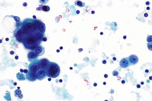
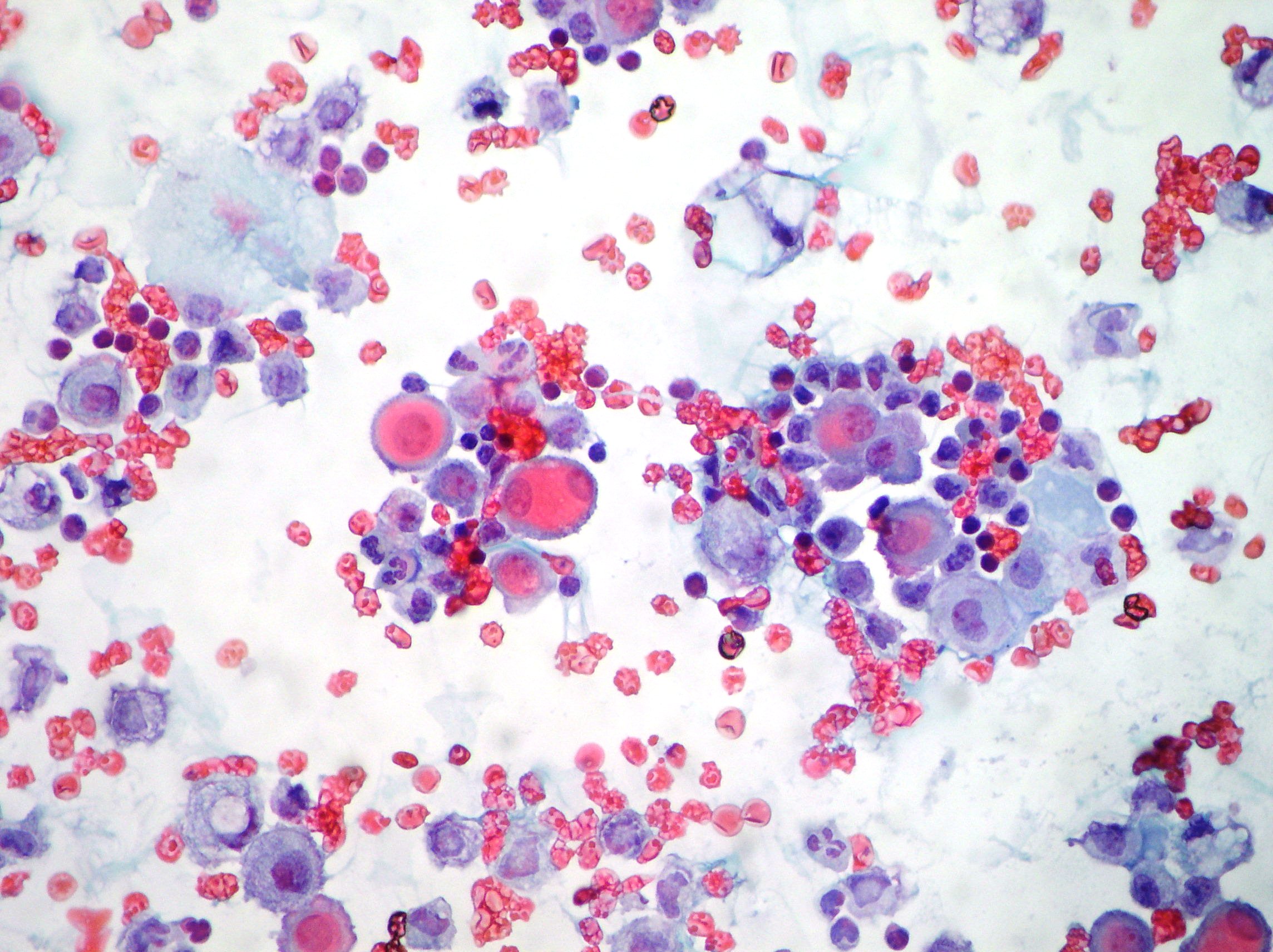
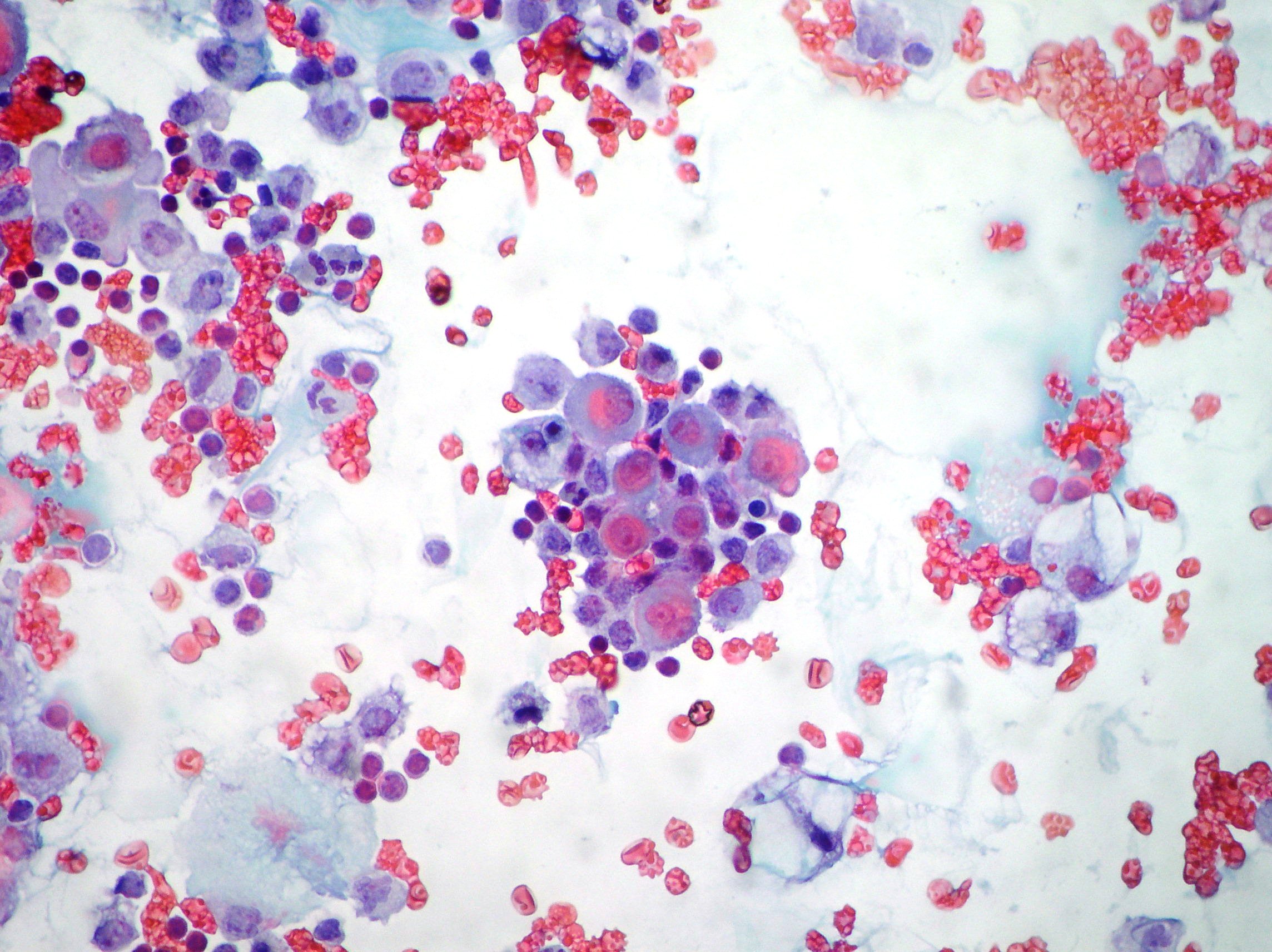
Reactive pleural effusion showing mesothelial cells lymphocytes neutrophils and macrophages. The fluid can accumulate quickly if the mesothelial cells fail to function resulting in an unhealthy collection of fluids in the chest cavity.


Effusions Cytopathology Cellnetpathology

Numerous reactive mesothelial cells were present in only 12 of specimens examined.
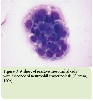
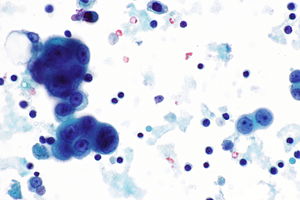
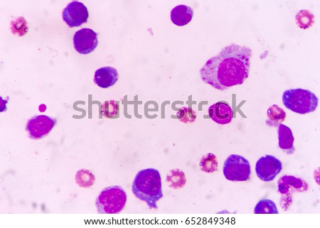
Mesothelial cells in pleural fluid. Reactive mesothelial cells present in a background of abundant lymphocytes. This has a large ddx. This condition can be due to the presence of a bacterial viral or fungal infection.

It can also be the result of trauma or the presence of metastatic tumor. Reactive mesothelial cells reactive mesothelial cells in pleural fluid reactive mesothelial cells are found when there is infection or inflammation present in a body cavity. Pleural effusion mesothelial cells pleural effusion.

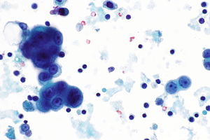
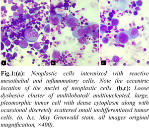
Negative for malignant cells. Pleural fluid right thoracentesis. Neoplastic transformation of mesothelial cells results in malignant mesothelioma an aggressive tumor especially the pleura.

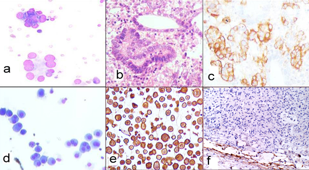
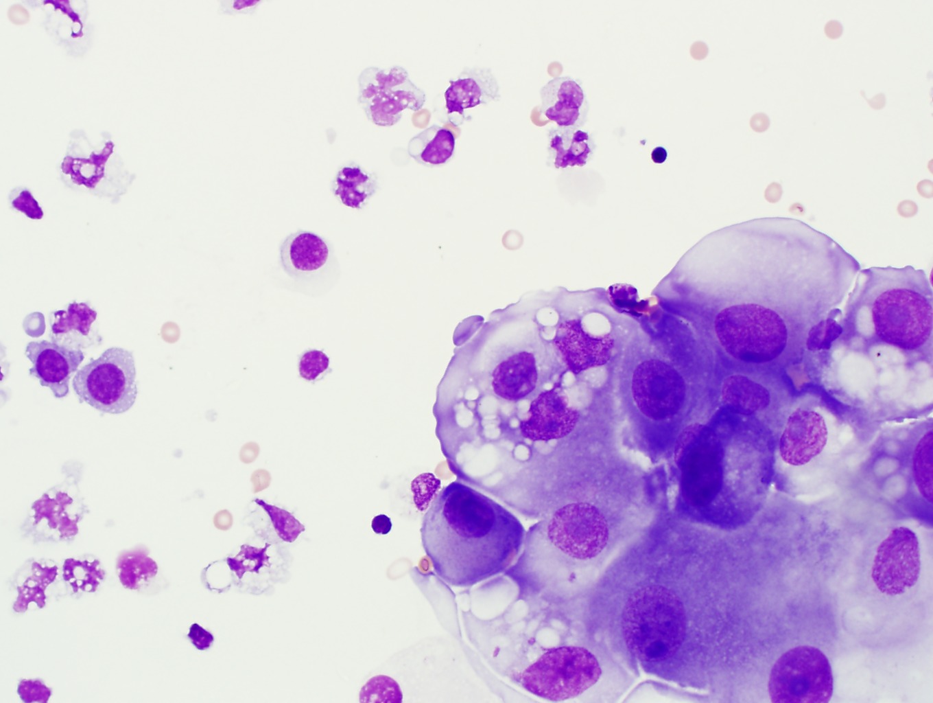
Immunocytochemistry test resulted positive for calretinin marker antibody papanicolaou x100 x200 mesothelial cells pleuric mesothelioma. Numerous mesothelial cells are seen in this pleural fluid from a dog with a transudative effusion with concurrent diapedesis of red blood cells or hemorrhage. Mesothelial cells are found in variable numbers in most effusions but their presence at greater than 5 of total nucleated cells makes a diagnosis of tb less likely.

This is known as pleural effusion. Reactive mesothelial cells can be found when there is an infection or an inflammatory response present in a body cavity. Actively dividing mesothelial cells can mimic an adenocarcinoma.

Pleural fluid for total white blood cell wbc count and differential cell count should be sent in an anticoagulated tube. A patient may develop shortness of breath and vague chest pains while the fluid experiences a buildup in the chest cavity these are two of the most common. Eighty five samples of pleural fluid obtained from 76 patients with biopsy proven tuberculous pleurisy were examined cytologically.

Specific diagnoses benign eosinophilic pleuritis general. Mesothelial cells in pleural fluid. Additional sampling should be considered within the clinical context.

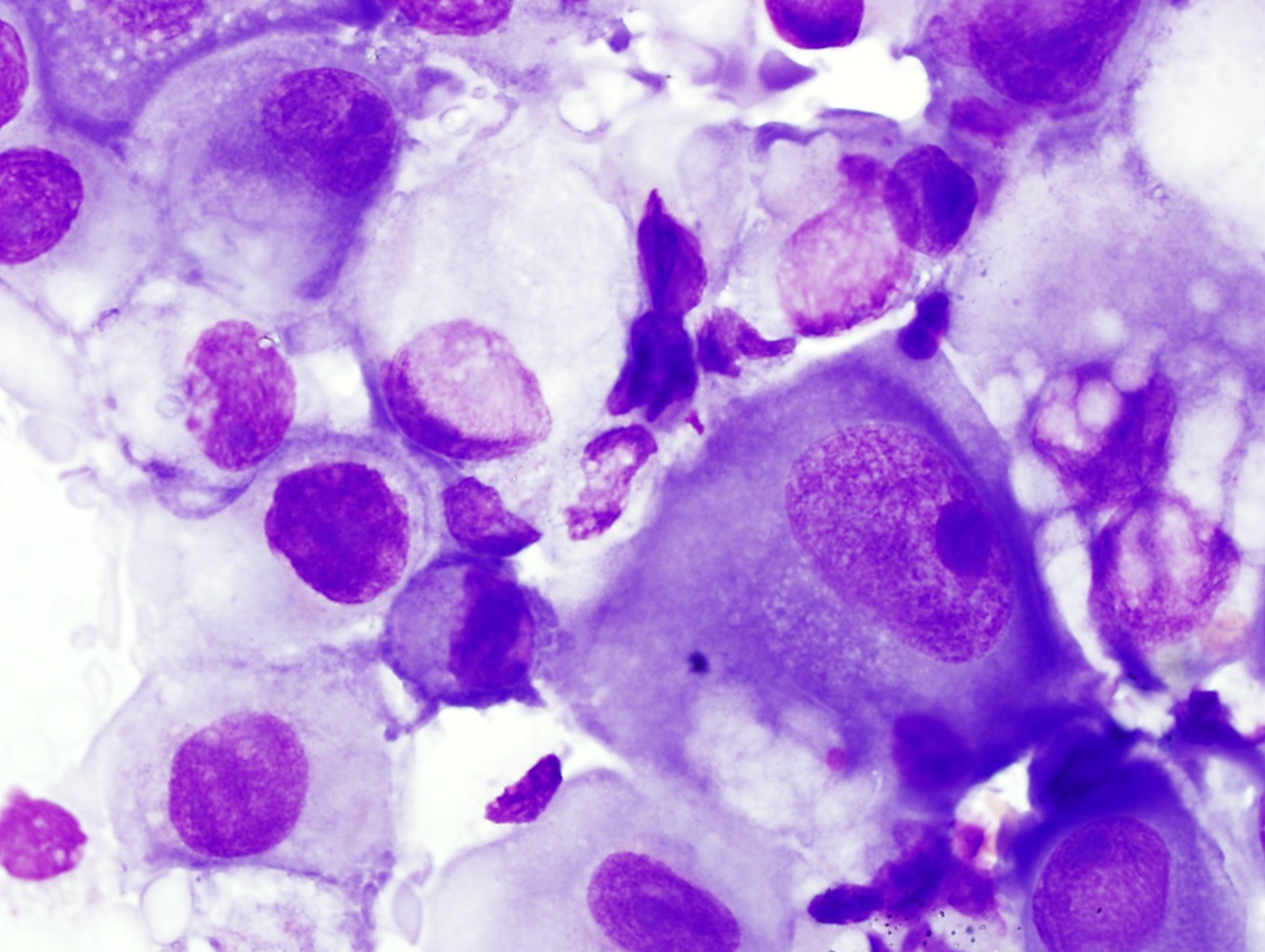
The mesothelial cells have central round nuclei with a moderate amount of light purple cytoplasm and a corona or fringe to the cytoplasmic borders. In contrast 653 of pleural fluid aspirates obtained from a control group of pati. There are certain cells that line the pleura the thin double layered lining which covers the lungs chest wall and diaphragm which are known as mesothelial cellsother than the pleura mesothelial cells also form a lining around the heart pericardium and the internal surface of the abdomen peritoneum.

Use of pleural fluid n. Trauma with air in the pleural cavity.


Http Www Cap Org Apps Docs Committees Hematology Educational Activities 2009 Cmb Pdf


Effusions Cytopathology Cellnetpathology


Pleural Fluid Smear Malignant Mesothelial Cells Lymphocytes Mgg Download Scientific Diagram
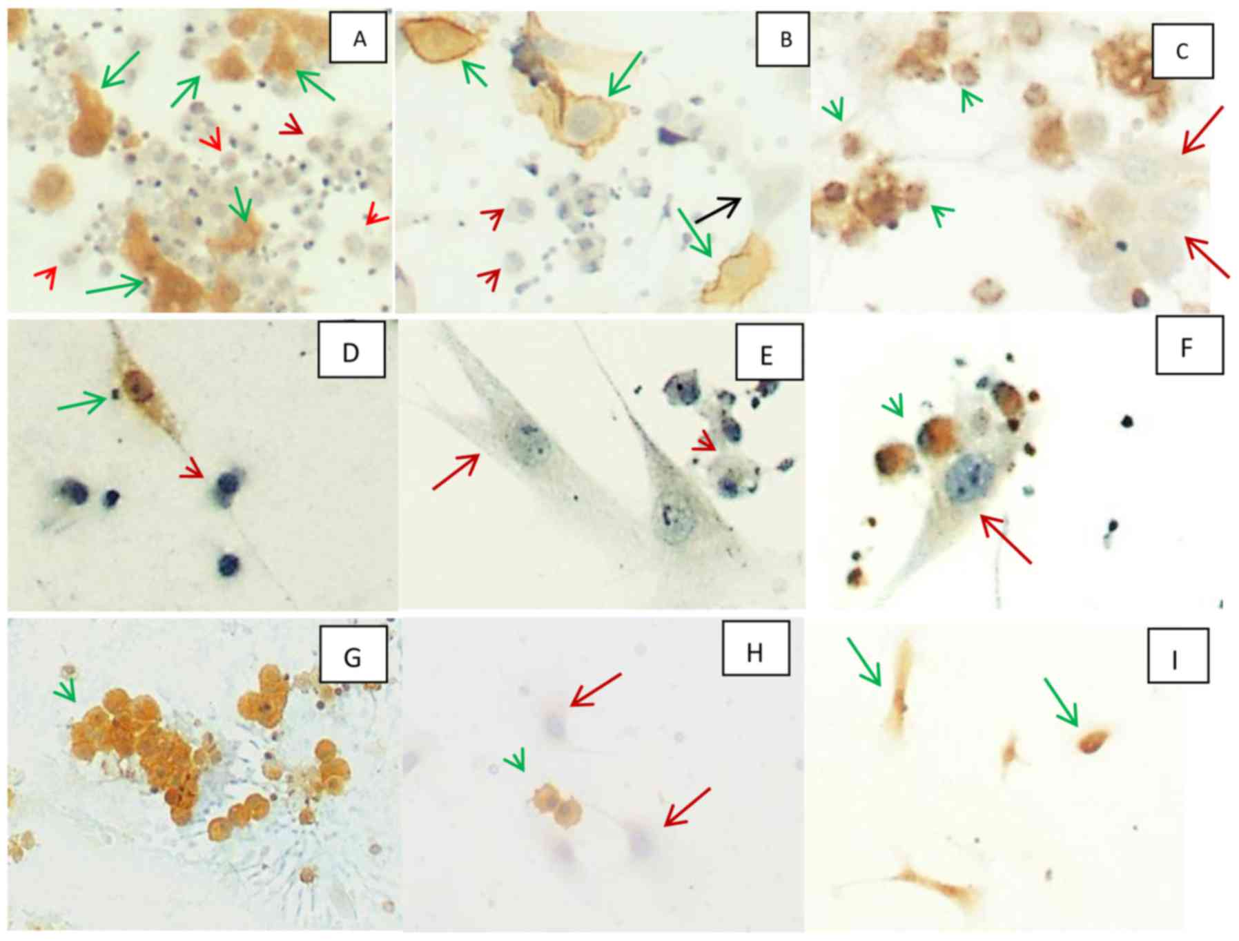


A Panel Of Markers For Identification Of Malignant And Non Malignant Cells In Culture From Effusions


View Image


Hjcam Iatrikh Zwwn Syntrofias Hellenic Journal Of Companion Animal Medicine Volume 6 Issue 1 2017 Pleural Effusion In The Cat A Focus On Laboratory Diagnosis


Http Www Cap Org Apps Docs Committees Hematology Educational Activities 2009 Cmb Pdf
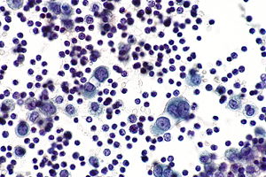
Reactive Mesothelial Cells In Pleural Fluid

It can also be the result of trauma or the presence of metastatic tumor. Additional sampling should be considered within the clinical context.


Home

Mesothelial cells are found in variable numbers in most effusions but their presence at greater than 5 of total nucleated cells makes a diagnosis of tb less likely.

Reactive mesothelial cells in pleural fluid. This has a large ddx. Trauma with air in the pleural cavity. In contrast 653 of pleural fluid aspirates obtained from a control group of pati.

Reactive mesothelial cells can be found when there is an infection or an inflammatory response present in a body cavity. Reactive mesothelial cells may sometimes be sampled in mediastinal fine needle aspirates and should not be mistaken as thymoma cells. Benign mesothelial cells tend to arrange in monolayered sheets with little nuclear overlapping fig.

Reactive mesothelial cells present in a background of abundant lymphocytes. The mesothelial cells have central round nuclei with a moderate amount of light purple cytoplasm and a corona or fringe to the cytoplasmic borders. This condition can be caused by the presence of bacterial viral or fungal.

Specific diagnoses benign eosinophilic pleuritis general. Negative for malignant cells. There are certain cells that line the pleura the thin double layered lining which covers the lungs chest wall and diaphragm which are known as mesothelial cellsother than the pleura mesothelial cells also form a lining around the heart pericardium and the internal surface of the abdomen peritoneum.

They contain ovoid nuclei fine chromatin delicate nuclear membrane small nucleoli and a moderate. It can also be the result of trauma or the presence of metastatic cancer. Reactive mesothelial cells in pleural fluid reactive mesothelial cells are found when there is infection or inflammation present in a body cavity.

Numerous reactive mesothelial cells were present in only 12 of specimens examined. Reactive mesothelial cells tend to come in. Reactive pleural effusion showing acute and chronic cells normal mesothelial cells and alveolar macrophages in aggregates and dispersed cells with rounded nuclei and vacuolated cytoplasm.

Mesothelial cells in pleural fluid. Use of pleural fluid n. Papanicolaou x100 breast adenocarcinoma cells in pleural effusion.

Numerous mesothelial cells are seen in this pleural fluid from a dog with a transudative effusion with concurrent diapedesis of red blood cells or hemorrhage. This condition can be due to the presence of a bacterial viral or fungal infection. Pleural fluid right thoracentesis.

Eighty five samples of pleural fluid obtained from 76 patients with biopsy proven tuberculous pleurisy were examined cytologically.


Http Oaji Net Pdf Html N 2015 1770 1438064825 Pdf


Pleural Fluid Mast Cells 2


Cytologic Differential Diagnosis Among Reactive Mesothelial Cells Malignant Mesothelioma And Adenocarcinoma Kitazume 2000 Cancer Cytopathology Wiley Online Library


Figure 3 Peritoneal Fluid From A Dog With A Dog With A Ruptured Mucocele Eclinpath


Https Onlinelibrary Wiley Com Doi Pdf 10 1002 Dc 20938


Effusions


Serous Effusions Basicmedical Key

Mesothelial Cells In Pleural Fluid Cytology

The diagnostic significance of the cytologic study of the fluid may be attributable to the fact that the cell population present in the sediment is representative of a. Numerous mesothelial cells are seen in this pleural fluid from a dog with a transudative effusion with concurrent diapedesis of red blood cells or hemorrhage.


Jcdr Adenocarcinoma Immunocytochemistry Reactive Mesothelial Cells Serous Effusions

Berger and stefan e.

Mesothelial cells in pleural fluid cytology. 20 of 119 of patients with malignancy involving the pleura but a negative pleural fluid cytology. An introduction to cytopathology is in the cytopathology article. Eighty five samples of mesothelial cells in pleural fluid from 76 patients with biopsy proven tuberculous pleurisy were examined cytologically.

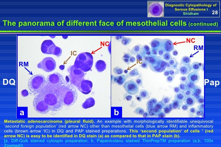
The morphological evaluation of cytological specimens from body cavity fluids presents difficulties in the differential diagnosis between benign reactive mesothelial rm cells and adenocarcinoma ac or malignant mesothelioma mm. Many reactive mesothelial cells were present in only 12 of samples analyzed. Actively dividing mesothelial cells can mimic an adenocarcinoma.

Pambuccian cytology of metastatic cervical squamous cell carcinoma in pleural fluid. Both non malignant and malignant causes of effusion can be identified by the relatively non invasive technique of pleural fluid cytologywith this basis the present study on cytology of pleural fluids was taken up. It deals with pericardial fluid peritoneal fluid and pleural fluid.

Gamez jose jessurun michael j. There are certain cells that line the pleura the thin double layered lining which covers the lungs chest wall and diaphragm which are known as mesothelial cellsother than the pleura mesothelial cells also form a lining around the heart pericardium and the internal surface of the abdomen peritoneum. Mesothelial cytopathology is a large part of cytopathology.

Mesothelial cells in pleural fluid. The aim of our study was to investigate whether a panel of five dif. The mesothelial cells have central round nuclei with a moderate amount of light purple cytoplasm and a corona or fringe to the cytoplasmic borders.

The article deals with cytopathology specimens from spaces lined with mesothelium ie. In contrast 653 of pleural fluid aspirates from a control group of patients with congestive heart failure contained. Report of a case confirmed by human papillomavirus typing diagnostic cytopathology 37 5 381 387 2009.

Cytology of mesothelial cells round with indistinct cell membranes and fuzzy borders single or clumped with so called windows between cells large round or elliptical nucleus often centrally placed with a single prominent nucleolus may contain two or multiple nuclei cytoplasm is green with papanicolaou and blue with mgg.


A Panel Of Markers For Identification Of Malignant And Non Malignant Cells In Culture From Effusions


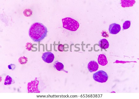
Benign Pleural Fluid Cytology Acute Inflammatory Stock Photo Edit Now 552969733


Shutterstock Puzzlepix


Hjcam Iatrikh Zwwn Syntrofias Hellenic Journal Of Companion Animal Medicine Volume 6 Issue 1 2017 Pleural Effusion In The Cat A Focus On Laboratory Diagnosis


The Value Of Cytology And Pleural Biopsy In The Differential Diagnostic Of Nonspecific Pleural Effusions


Http Iap Ad Org Documents Archieve Congress 25thcongress Workshop First 20day Body 20fluids 20cytology Pdf


Utility Of Cell Block To Detect Malignancy In Fluid Cytology Adjunct Or Necessity Dey S Nag D Nandi A Bandyopadhyay R J Can Res Ther

Reactive Mesothelial Cells In Pleural Fluid Cytology

Small cell carcinoma isolated cells clusters and chains round or angular nuclei molding may show elongation and spindling nuclear molding. An introduction to cytopathology is in the cytopathology article.


Home

Ihc and pap cytology are more reliable pleural fluid from a 73 yo man.

Reactive mesothelial cells in pleural fluid cytology. Reactive mesothelial cells reactive mesothelial cells in pleural fluid reactive mesothelial cells are found when there is infection or inflammation present in a body cavity. Reactive mesothelial cells may sometimes be sampled in mediastinal fine needle aspirates and should not be mistaken as thymoma cells. It deals with pericardial fluid peritoneal fluid and pleural fluid.

Fna lung pleural fluid. Although cytological examination helps in diagnosis of malignancy in serous effusion at times it is difficult to differentiate atypical reactive mesothelial cells from adenocarcinoma ac cells. Mesothelial cytopathology is a large part of cytopathology.

Neg in reactive mesothelial cells in 4 of cases. To resolve this problem various ancillary methods have been used. Further work up the tumor cells were also positive for wt1 calretinin figure 1c1 and andc2 c2 p53 and cytokeratin 56 and stained negatively for pax 8 berep4 desmin cea and ttf 1.

Actively dividing mesothelial cells can mimic an adenocarcinoma. Junko ueda takako iwata midori ono mutsuo takahashi comparison of three cytologic preparation methods and immunocytochemistries to distinguish adenocarcinoma cells from reactive mesothelial cells in serous effusion diagnostic cytopathology 101002dc20391 34 1 6 10 2005. Immunocytochemistry icc is one such commonly used technique in which various panel of antibodies has been tried.

20 of 119 of patients with malignancy involving the pleura but a negative pleural fluid cytology. They contain ovoid nuclei fine chromatin delicate nuclear membrane small nucleoli and a moderate. The reactive mesothelial proliferations form smaller uniform less complex groups compared to malignant mesothelial proliferations.

To evaluate the use of a panel of markers to differentiate adenocarcinoma and the reactiveinflammatory process in fluid cytology we stained 29 formalin fixed paraffin embedded cell blocks of effusion fluid from patients with metastatic adenocarcinoma and 24 cell blocks from patients with benign effusion with mucicarmine and antibodies to carcinoembryonic antigen cea b723 and calretinin. Benign mesothelial cells tend to arrange in monolayered sheets with little nuclear overlapping fig. The article deals with cytopathology specimens from spaces lined with mesothelium ie.

Neoplastic transformation of mesothelial cells results in malignant mesothelioma an aggressive tumor especially the pleura.


Effusion Cytomorphology Of Rhabdomyosarcoma A Rare Case Of Primary Mediastinal Rhabdomyosarcoma With Superior Vena Cava Obstruction And Bilateral Pleural Effusion


01 Presentation I Vs 8 55mb 3 28 08 Pps


Https Www Rcpath Org Asset Ed8cdd8d 8d04 4b82 Ad48d585e2f023be


Body Cavity Effusions Are Among The Most Commonly Received


Shutterstock Puzzlepix


Https Www Sciencedirect Com Science Article Pii S1877050913010909 Pdf Md5 Dcd46a4816e9d057d4912a8943bc10d6 Pid 1 S2 0 S1877050913010909 Main Pdf


Mesothelioma In A Pleural Fluid Specimen Atypical Mesothelial Cells Download Scientific Diagram