Tag Archives: reactive mesothelial cells in pleural fluid pathology outlines

Reactive Mesothelial Cells In Pleural Fluid Pathology Outlines

It may also include repopulation from free floating mesothelial cells or possibly. Ecadherin is an adhesion protein that is specifically expressed in cells of epithelial lineage.


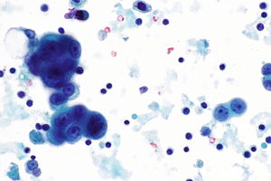
Mesothelioma Pleural Effusion

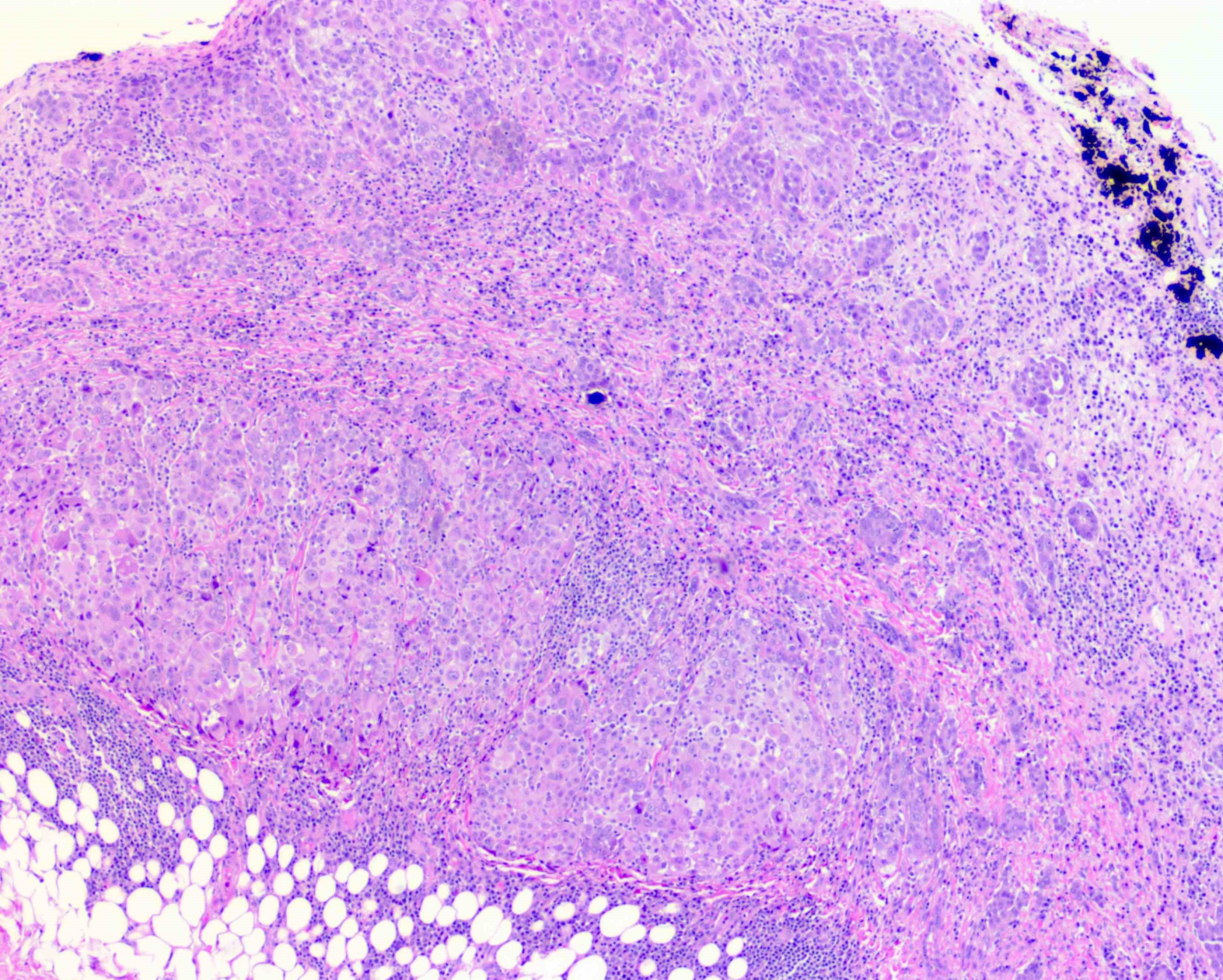
Reactive mesothelial proliferation 6 8 on morphology signs of malignancy include invasion for instance in the adipose tissue lung and skeletal muscle and the keratin really can be helpful to highlight the invasion of the neoplastic cells.

Reactive mesothelial cells in pleural fluid pathology outlines. The mesothelial cell clone hbme1 is an antibody against cultured mesothelial cells and recognizes an antigen on the microvillus surface. Weiss in modern surgical pathology second edition 2009. Mesothelial hyperplasia represents a normal reaction to injury.

Pleural peritoneal and pericardial fluids were prepared using the cytyc thin prep processor. Specific diagnoses benign eosinophilic pleuritis general. Ecadherin is an adhesion protein that is specifically expressed in cells of epithelial lineage.

Trauma with air in the pleural cavity. Reactive proliferation of stromal myofibroblasts often associated with adjacent linear arrangement of mesothelial cells chronic inflammation surface fibrin deposition and occasionally entrapped fat associated with various conditions including luteinized thecomas am j surg pathol 1994181. This has a large ddx.

In this study antiecadherin antibodies were used to identify and distinguish carcinoma cells from reactive mesothelial cells. The healing of disrupted serosa includes the multiplication and migration of mesothelial cells from the edges of the injured area. Reactive mesothelial cells present in a background of abundant lymphocytes.

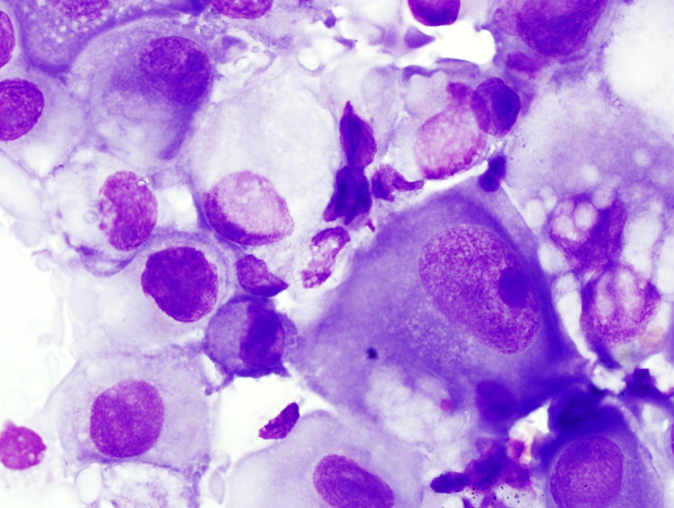
The distinction between benign reactive mesothelial cells and well differentiated carcinoma can be difficult in pleural peritoneal and especially pericardial fluids. Mamatha chivukula david j. Hbme1 has been used as a part of the panel of icc to distinguish adenocarcinoma from mesothelial cells.

Dabbs in diagnostic immunohistochemistry third edition 2011. Additional sampling should be considered within the clinical context. Pleural fluid right thoracentesis.

Negative for malignant cells.


Pdf Peritoneal Washing Cytology


Pathology Outlines Mesothelioma Epithelioid


Mesothelioma Vs Reactive Mesothelial Cells Cytology Creative Art


Do7vxfhb7u 2nm


Outlines In Pathology John H Sinard Md Phd


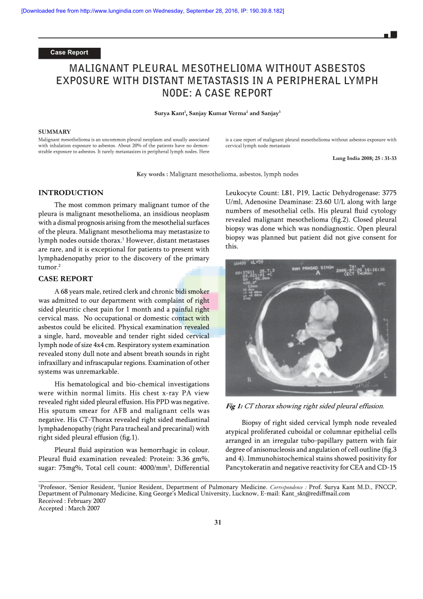
Pdf Malignant Pleural Mesothelioma Without Asbestos Exposure With Distant Metastasis In A Peripheral Lymph Node A Case Report


Cytology Clinical Pathology And Procedures Merck Veterinary Manual

Reactive Mesothelial Cells In Pleural Fluid

It can also be the result of trauma or the presence of metastatic tumor. Additional sampling should be considered within the clinical context.


Home

Mesothelial cells are found in variable numbers in most effusions but their presence at greater than 5 of total nucleated cells makes a diagnosis of tb less likely.

Reactive mesothelial cells in pleural fluid. This has a large ddx. Trauma with air in the pleural cavity. In contrast 653 of pleural fluid aspirates obtained from a control group of pati.

Reactive mesothelial cells can be found when there is an infection or an inflammatory response present in a body cavity. Reactive mesothelial cells may sometimes be sampled in mediastinal fine needle aspirates and should not be mistaken as thymoma cells. Benign mesothelial cells tend to arrange in monolayered sheets with little nuclear overlapping fig.

Reactive mesothelial cells present in a background of abundant lymphocytes. The mesothelial cells have central round nuclei with a moderate amount of light purple cytoplasm and a corona or fringe to the cytoplasmic borders. This condition can be caused by the presence of bacterial viral or fungal.

Specific diagnoses benign eosinophilic pleuritis general. Negative for malignant cells. There are certain cells that line the pleura the thin double layered lining which covers the lungs chest wall and diaphragm which are known as mesothelial cellsother than the pleura mesothelial cells also form a lining around the heart pericardium and the internal surface of the abdomen peritoneum.

They contain ovoid nuclei fine chromatin delicate nuclear membrane small nucleoli and a moderate. It can also be the result of trauma or the presence of metastatic cancer. Reactive mesothelial cells in pleural fluid reactive mesothelial cells are found when there is infection or inflammation present in a body cavity.

Numerous reactive mesothelial cells were present in only 12 of specimens examined. Reactive mesothelial cells tend to come in. Reactive pleural effusion showing acute and chronic cells normal mesothelial cells and alveolar macrophages in aggregates and dispersed cells with rounded nuclei and vacuolated cytoplasm.

Mesothelial cells in pleural fluid. Use of pleural fluid n. Papanicolaou x100 breast adenocarcinoma cells in pleural effusion.

Numerous mesothelial cells are seen in this pleural fluid from a dog with a transudative effusion with concurrent diapedesis of red blood cells or hemorrhage. This condition can be due to the presence of a bacterial viral or fungal infection. Pleural fluid right thoracentesis.

Eighty five samples of pleural fluid obtained from 76 patients with biopsy proven tuberculous pleurisy were examined cytologically.


Http Oaji Net Pdf Html N 2015 1770 1438064825 Pdf


Pleural Fluid Mast Cells 2


Cytologic Differential Diagnosis Among Reactive Mesothelial Cells Malignant Mesothelioma And Adenocarcinoma Kitazume 2000 Cancer Cytopathology Wiley Online Library


Figure 3 Peritoneal Fluid From A Dog With A Dog With A Ruptured Mucocele Eclinpath


Https Onlinelibrary Wiley Com Doi Pdf 10 1002 Dc 20938


Effusions


Serous Effusions Basicmedical Key